Calcium »
PDB 4cgt-4cud »
4cj1 »
Calcium in PDB 4cj1: Crystal Structure of Celd in Complex with Affitin H3
Enzymatic activity of Crystal Structure of Celd in Complex with Affitin H3
All present enzymatic activity of Crystal Structure of Celd in Complex with Affitin H3:
3.2.1.4;
3.2.1.4;
Protein crystallography data
The structure of Crystal Structure of Celd in Complex with Affitin H3, PDB code: 4cj1
was solved by
A.Correa,
S.Pacheco,
A.E.Mechaly,
G.Obal,
G.Behar,
B.Mouratou,
P.Oppezzo,
P.M.Alzari,
F.Pecorari,
with X-Ray Crystallography technique. A brief refinement statistics is given in the table below:
| Resolution Low / High (Å) | 46.83 / 1.63 |
| Space group | P 21 21 21 |
| Cell size a, b, c (Å), α, β, γ (°) | 74.422, 97.736, 106.580, 90.00, 90.00, 90.00 |
| R / Rfree (%) | 10.608 / 14.374 |
Other elements in 4cj1:
The structure of Crystal Structure of Celd in Complex with Affitin H3 also contains other interesting chemical elements:
| Zinc | (Zn) | 1 atom |
Calcium Binding Sites:
The binding sites of Calcium atom in the Crystal Structure of Celd in Complex with Affitin H3
(pdb code 4cj1). This binding sites where shown within
5.0 Angstroms radius around Calcium atom.
In total 3 binding sites of Calcium where determined in the Crystal Structure of Celd in Complex with Affitin H3, PDB code: 4cj1:
Jump to Calcium binding site number: 1; 2; 3;
In total 3 binding sites of Calcium where determined in the Crystal Structure of Celd in Complex with Affitin H3, PDB code: 4cj1:
Jump to Calcium binding site number: 1; 2; 3;
Calcium binding site 1 out of 3 in 4cj1
Go back to
Calcium binding site 1 out
of 3 in the Crystal Structure of Celd in Complex with Affitin H3

Mono view
Stereo pair view
Mono view
Stereo pair view
A full contact list of Calcium with other atoms in the Ca binding
site number 1 of Crystal Structure of Celd in Complex with Affitin H3 within 5.0Å range:
|
Calcium binding site 2 out of 3 in 4cj1
Go back to
Calcium binding site 2 out
of 3 in the Crystal Structure of Celd in Complex with Affitin H3

Mono view

Stereo pair view
Mono view
Stereo pair view
A full contact list of Calcium with other atoms in the Ca binding
site number 2 of Crystal Structure of Celd in Complex with Affitin H3 within 5.0Å range:
|
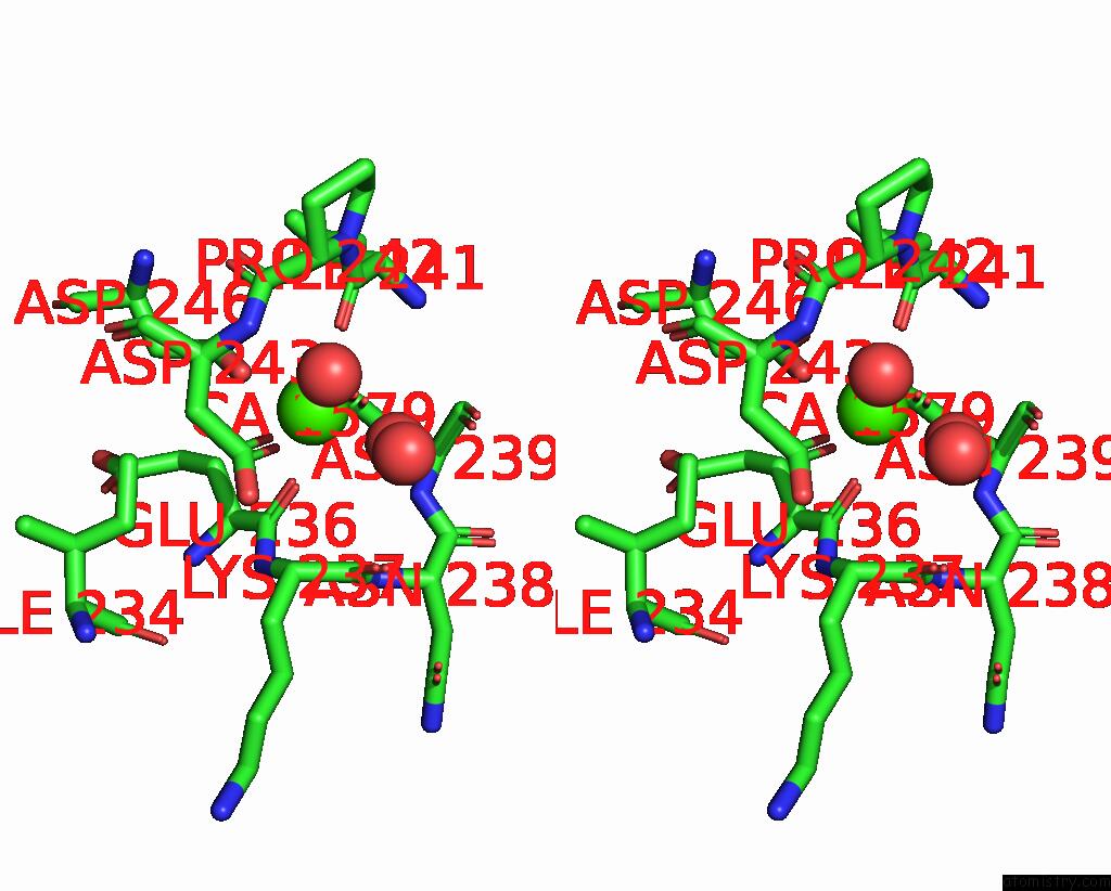
Calcium binding site 3 out of 3 in 4cj1
Go back to
Calcium binding site 3 out
of 3 in the Crystal Structure of Celd in Complex with Affitin H3

Mono view

Stereo pair view
Mono view
Stereo pair view
A full contact list of Calcium with other atoms in the Ca binding
site number 3 of Crystal Structure of Celd in Complex with Affitin H3 within 5.0Å range:
|
Reference:
A.Correa,
S.Pacheco,
A.E.Mechaly,
G.Obal,
G.Behar,
B.Mouratou,
P.Oppezzo,
P.M.Alzari,
F.Pecorari.
Potent and Specific Inhibition of Glycosidases By Small Artificial Binding Proteins (Affitins) Plos One V. 9 97438 2014.
ISSN: ISSN 1932-6203
PubMed: 24823716
DOI: 10.1371/JOURNAL.PONE.0097438
Page generated: Tue Jul 8 19:14:05 2025
ISSN: ISSN 1932-6203
PubMed: 24823716
DOI: 10.1371/JOURNAL.PONE.0097438
Last articles
Mg in 5SHYMg in 5SHV
Mg in 5SHU
Mg in 5SHT
Mg in 5SHS
Mg in 5SHR
Mg in 5SHQ
Mg in 5SHP
Mg in 5SHN
Mg in 5SHO