Calcium »
PDB 4cgt-4cud »
4cp0 »
Calcium in PDB 4cp0: Crystal Structure of Epithelial Adhesin 9 A Domain (EPA9A) From Candida Glabrata in Complex with Lactose
Protein crystallography data
The structure of Crystal Structure of Epithelial Adhesin 9 A Domain (EPA9A) From Candida Glabrata in Complex with Lactose, PDB code: 4cp0
was solved by
M.Kock,
M.Maestre-Reyna,
R.Diderrich,
H.-U.Moesch,
L.-O.Essen,
with X-Ray Crystallography technique. A brief refinement statistics is given in the table below:
| Resolution Low / High (Å) | 41.23 / 2.15 |
| Space group | P 31 2 1 |
| Cell size a, b, c (Å), α, β, γ (°) | 66.580, 66.580, 117.950, 90.00, 90.00, 120.00 |
| R / Rfree (%) | 16.391 / 19.381 |
Other elements in 4cp0:
The structure of Crystal Structure of Epithelial Adhesin 9 A Domain (EPA9A) From Candida Glabrata in Complex with Lactose also contains other interesting chemical elements:
| Chlorine | (Cl) | 1 atom |
Calcium Binding Sites:
The binding sites of Calcium atom in the Crystal Structure of Epithelial Adhesin 9 A Domain (EPA9A) From Candida Glabrata in Complex with Lactose
(pdb code 4cp0). This binding sites where shown within
5.0 Angstroms radius around Calcium atom.
In total only one binding site of Calcium was determined in the Crystal Structure of Epithelial Adhesin 9 A Domain (EPA9A) From Candida Glabrata in Complex with Lactose, PDB code: 4cp0:
In total only one binding site of Calcium was determined in the Crystal Structure of Epithelial Adhesin 9 A Domain (EPA9A) From Candida Glabrata in Complex with Lactose, PDB code: 4cp0:
Calcium binding site 1 out of 1 in 4cp0
Go back to
Calcium binding site 1 out
of 1 in the Crystal Structure of Epithelial Adhesin 9 A Domain (EPA9A) From Candida Glabrata in Complex with Lactose
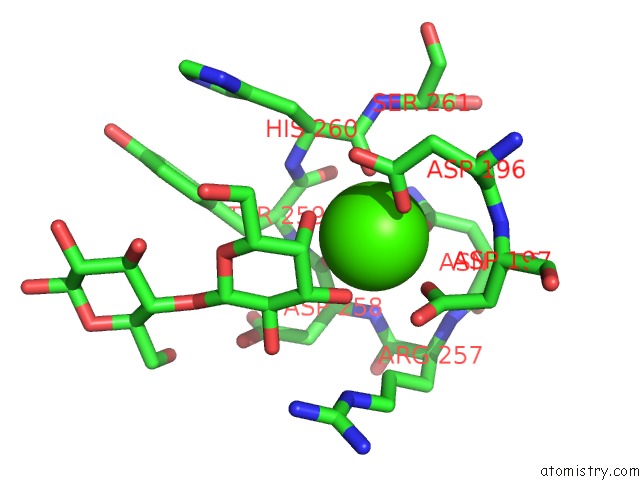
Mono view
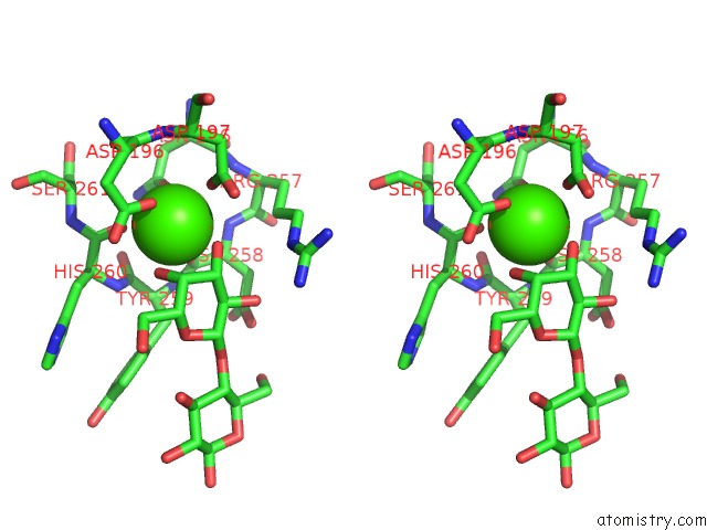
Stereo pair view
Mono view
Stereo pair view
A full contact list of Calcium with other atoms in the Ca binding
site number 1 of Crystal Structure of Epithelial Adhesin 9 A Domain (EPA9A) From Candida Glabrata in Complex with Lactose within 5.0Å range:
|
Reference:
R.Diderrich,
M.Kock,
M.Maestre-Reyna,
S.Rupp,
L.-O.Essen,
H.-U.Moesch.
The Structure and Function of the Epithelial Adhesins (Working) To Be Published.
Page generated: Tue Jul 8 19:15:24 2025
Last articles
Mg in 5MMJMg in 5MRA
Mg in 5MTV
Mg in 5MS0
Mg in 5MRU
Mg in 5MQJ
Mg in 5MQW
Mg in 5MQT
Mg in 5MQL
Mg in 5MQ1