Calcium »
PDB 4dtm-4ec5 »
4e55 »
Calcium in PDB 4e55: Crystal Structure of Spacer Removed Cephalosporin Acylase Mutant
Enzymatic activity of Crystal Structure of Spacer Removed Cephalosporin Acylase Mutant
All present enzymatic activity of Crystal Structure of Spacer Removed Cephalosporin Acylase Mutant:
3.5.1.93;
3.5.1.93;
Protein crystallography data
The structure of Crystal Structure of Spacer Removed Cephalosporin Acylase Mutant, PDB code: 4e55
was solved by
J.Yin,
Z.Zhang,
G.Wu,
X.Huang,
with X-Ray Crystallography technique. A brief refinement statistics is given in the table below:
| Resolution Low / High (Å) | 50.00 / 2.30 |
| Space group | P 41 |
| Cell size a, b, c (Å), α, β, γ (°) | 74.238, 74.238, 383.860, 90.00, 90.00, 90.00 |
| R / Rfree (%) | 17.1 / 19.9 |
Calcium Binding Sites:
The binding sites of Calcium atom in the Crystal Structure of Spacer Removed Cephalosporin Acylase Mutant
(pdb code 4e55). This binding sites where shown within
5.0 Angstroms radius around Calcium atom.
In total 2 binding sites of Calcium where determined in the Crystal Structure of Spacer Removed Cephalosporin Acylase Mutant, PDB code: 4e55:
Jump to Calcium binding site number: 1; 2;
In total 2 binding sites of Calcium where determined in the Crystal Structure of Spacer Removed Cephalosporin Acylase Mutant, PDB code: 4e55:
Jump to Calcium binding site number: 1; 2;
Calcium binding site 1 out of 2 in 4e55
Go back to
Calcium binding site 1 out
of 2 in the Crystal Structure of Spacer Removed Cephalosporin Acylase Mutant

Mono view
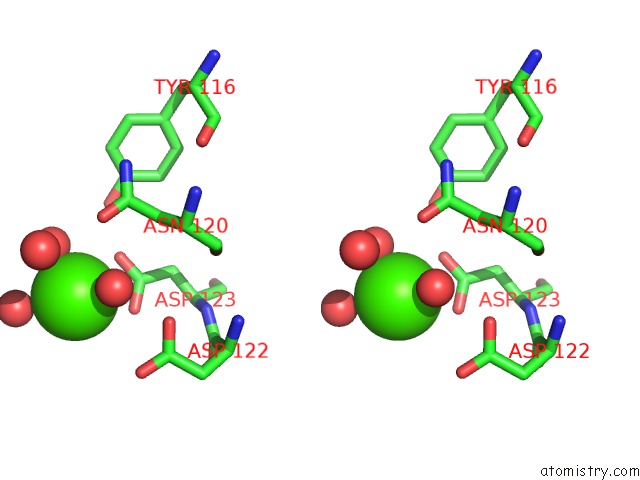
Stereo pair view
Mono view
Stereo pair view
A full contact list of Calcium with other atoms in the Ca binding
site number 1 of Crystal Structure of Spacer Removed Cephalosporin Acylase Mutant within 5.0Å range:
|
Calcium binding site 2 out of 2 in 4e55
Go back to
Calcium binding site 2 out
of 2 in the Crystal Structure of Spacer Removed Cephalosporin Acylase Mutant

Mono view

Stereo pair view
Mono view
Stereo pair view
A full contact list of Calcium with other atoms in the Ca binding
site number 2 of Crystal Structure of Spacer Removed Cephalosporin Acylase Mutant within 5.0Å range:
|
Reference:
J.Yin,
Z.Zhang,
G.Wu,
X.Huang.
Spacer Shortened Cephalosporin Acylase To Be Published.
Page generated: Tue Jul 8 19:39:46 2025
Last articles
Mg in 6Y1DMg in 6Y1C
Mg in 6Y1B
Mg in 6Y15
Mg in 6Y18
Mg in 6XZ7
Mg in 6XU1
Mg in 6Y0Z
Mg in 6Y0T
Mg in 6Y0Y