Calcium »
PDB 4ecg-4enz »
4ee2 »
Calcium in PDB 4ee2: Crystal Structure of Anthrax Protective Antigen K446M Mutant to 1.91-A Resolution
Protein crystallography data
The structure of Crystal Structure of Anthrax Protective Antigen K446M Mutant to 1.91-A Resolution, PDB code: 4ee2
was solved by
A.F.Kintzer,
B.A.Krantz,
with X-Ray Crystallography technique. A brief refinement statistics is given in the table below:
| Resolution Low / High (Å) | 40.69 / 1.91 |
| Space group | P 21 21 21 |
| Cell size a, b, c (Å), α, β, γ (°) | 71.216, 93.512, 116.918, 90.00, 90.00, 90.00 |
| R / Rfree (%) | 22.9 / 26.5 |
Calcium Binding Sites:
The binding sites of Calcium atom in the Crystal Structure of Anthrax Protective Antigen K446M Mutant to 1.91-A Resolution
(pdb code 4ee2). This binding sites where shown within
5.0 Angstroms radius around Calcium atom.
In total 2 binding sites of Calcium where determined in the Crystal Structure of Anthrax Protective Antigen K446M Mutant to 1.91-A Resolution, PDB code: 4ee2:
Jump to Calcium binding site number: 1; 2;
In total 2 binding sites of Calcium where determined in the Crystal Structure of Anthrax Protective Antigen K446M Mutant to 1.91-A Resolution, PDB code: 4ee2:
Jump to Calcium binding site number: 1; 2;
Calcium binding site 1 out of 2 in 4ee2
Go back to
Calcium binding site 1 out
of 2 in the Crystal Structure of Anthrax Protective Antigen K446M Mutant to 1.91-A Resolution
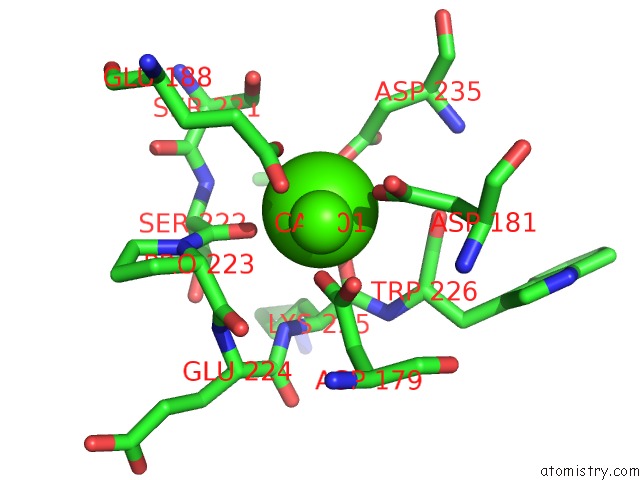
Mono view
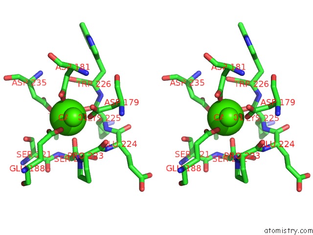
Stereo pair view
Mono view
Stereo pair view
A full contact list of Calcium with other atoms in the Ca binding
site number 1 of Crystal Structure of Anthrax Protective Antigen K446M Mutant to 1.91-A Resolution within 5.0Å range:
|
Calcium binding site 2 out of 2 in 4ee2
Go back to
Calcium binding site 2 out
of 2 in the Crystal Structure of Anthrax Protective Antigen K446M Mutant to 1.91-A Resolution
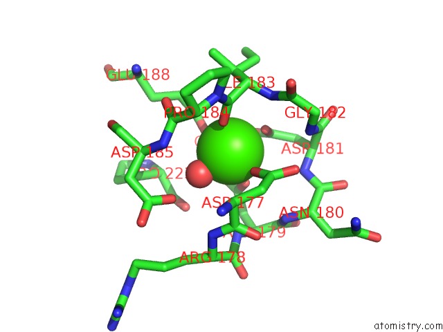
Mono view
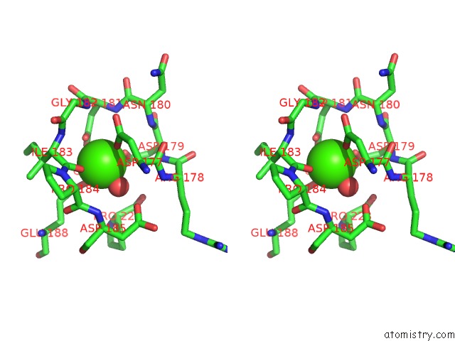
Stereo pair view
Mono view
Stereo pair view
A full contact list of Calcium with other atoms in the Ca binding
site number 2 of Crystal Structure of Anthrax Protective Antigen K446M Mutant to 1.91-A Resolution within 5.0Å range:
|
Reference:
A.F.Kintzer,
I.I.Tang,
A.K.Schawel,
M.J.Brown,
B.A.Krantz.
Anthrax Toxin Protective Antigen Integrates Poly-Gamma-D-Glutamate and pH Signals to Sense the Optimal Environment For Channel Formation. Proc.Natl.Acad.Sci.Usa V. 109 18378 2012.
ISSN: ISSN 0027-8424
PubMed: 23100533
DOI: 10.1073/PNAS.1208280109
Page generated: Tue Jul 8 19:45:35 2025
ISSN: ISSN 0027-8424
PubMed: 23100533
DOI: 10.1073/PNAS.1208280109
Last articles
Fe in 2YXOFe in 2YRS
Fe in 2YXC
Fe in 2YNM
Fe in 2YVJ
Fe in 2YP1
Fe in 2YU2
Fe in 2YU1
Fe in 2YQB
Fe in 2YOO