Calcium »
PDB 4eoy-4fb1 »
4eto »
Calcium in PDB 4eto: Structure of S100A4 in Complex with Non-Muscle Myosin-Iia Peptide
Protein crystallography data
The structure of Structure of S100A4 in Complex with Non-Muscle Myosin-Iia Peptide, PDB code: 4eto
was solved by
U.A.Ramagopal,
N.G.Dulyaninova,
P.R.Kumar,
S.C.Almo,
A.R.Bresnick,
Newyork Structural Genomics Research Consortium (Nysgrc),
Atoms-To-Animals: The Immune Function Network (Ifn),
with X-Ray Crystallography technique. A brief refinement statistics is given in the table below:
| Resolution Low / High (Å) | 45.99 / 1.54 |
| Space group | P 1 21 1 |
| Cell size a, b, c (Å), α, β, γ (°) | 30.279, 91.989, 32.864, 90.00, 112.60, 90.00 |
| R / Rfree (%) | 20.9 / 25.1 |
Calcium Binding Sites:
The binding sites of Calcium atom in the Structure of S100A4 in Complex with Non-Muscle Myosin-Iia Peptide
(pdb code 4eto). This binding sites where shown within
5.0 Angstroms radius around Calcium atom.
In total 4 binding sites of Calcium where determined in the Structure of S100A4 in Complex with Non-Muscle Myosin-Iia Peptide, PDB code: 4eto:
Jump to Calcium binding site number: 1; 2; 3; 4;
In total 4 binding sites of Calcium where determined in the Structure of S100A4 in Complex with Non-Muscle Myosin-Iia Peptide, PDB code: 4eto:
Jump to Calcium binding site number: 1; 2; 3; 4;
Calcium binding site 1 out of 4 in 4eto
Go back to
Calcium binding site 1 out
of 4 in the Structure of S100A4 in Complex with Non-Muscle Myosin-Iia Peptide

Mono view
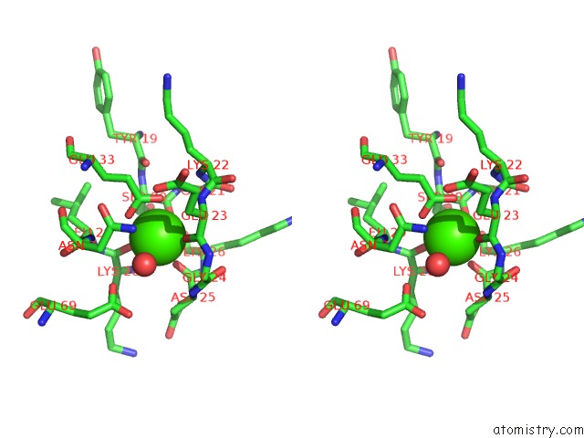
Stereo pair view
Mono view
Stereo pair view
A full contact list of Calcium with other atoms in the Ca binding
site number 1 of Structure of S100A4 in Complex with Non-Muscle Myosin-Iia Peptide within 5.0Å range:
|
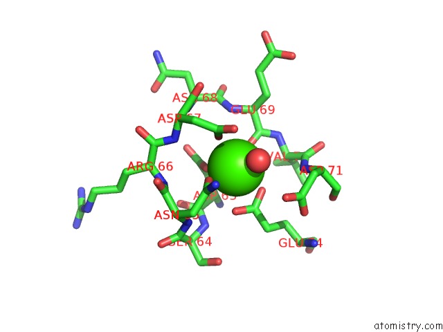
Calcium binding site 2 out of 4 in 4eto
Go back to
Calcium binding site 2 out
of 4 in the Structure of S100A4 in Complex with Non-Muscle Myosin-Iia Peptide

Mono view

Stereo pair view
Mono view
Stereo pair view
A full contact list of Calcium with other atoms in the Ca binding
site number 2 of Structure of S100A4 in Complex with Non-Muscle Myosin-Iia Peptide within 5.0Å range:
|
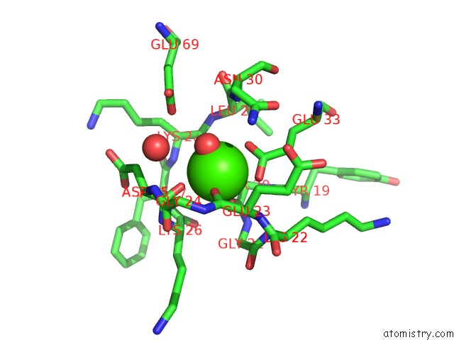
Calcium binding site 3 out of 4 in 4eto
Go back to
Calcium binding site 3 out
of 4 in the Structure of S100A4 in Complex with Non-Muscle Myosin-Iia Peptide
Mono view

Stereo pair view
Mono view
Stereo pair view
A full contact list of Calcium with other atoms in the Ca binding
site number 3 of Structure of S100A4 in Complex with Non-Muscle Myosin-Iia Peptide within 5.0Å range:
|
Calcium binding site 4 out of 4 in 4eto
Go back to
Calcium binding site 4 out
of 4 in the Structure of S100A4 in Complex with Non-Muscle Myosin-Iia Peptide
Mono view
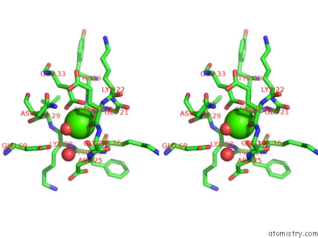
Stereo pair view
Mono view
Stereo pair view
A full contact list of Calcium with other atoms in the Ca binding
site number 4 of Structure of S100A4 in Complex with Non-Muscle Myosin-Iia Peptide within 5.0Å range:
|
Reference:
U.A.Ramagopal,
N.G.Dulyaninova,
P.R.Kumar,
S.C.Almo,
A.R.Bresnick.
Structure of S100A4 with Bound Peptide P To Be Published.
Page generated: Tue Jul 8 19:52:00 2025
Last articles
Mg in 6YBWMg in 6YKW
Mg in 6YKV
Mg in 6YKU
Mg in 6YKT
Mg in 6YKS
Mg in 6YKQ
Mg in 6YKO
Mg in 6YKN
Mg in 6YKL