Calcium »
PDB 4eoy-4fb1 »
4f7x »
Calcium in PDB 4f7x: Crystal Structure of Staphylococcal Nuclease Variant Delta+Phs I92A/L25A at Cryogenic Temperature
Enzymatic activity of Crystal Structure of Staphylococcal Nuclease Variant Delta+Phs I92A/L25A at Cryogenic Temperature
All present enzymatic activity of Crystal Structure of Staphylococcal Nuclease Variant Delta+Phs I92A/L25A at Cryogenic Temperature:
3.1.31.1;
3.1.31.1;
Protein crystallography data
The structure of Crystal Structure of Staphylococcal Nuclease Variant Delta+Phs I92A/L25A at Cryogenic Temperature, PDB code: 4f7x
was solved by
J.A.Caro,
I.Clark,
J.L.Schlessman,
A.Heroux,
B.Garcia-Moreno E.,
with X-Ray Crystallography technique. A brief refinement statistics is given in the table below:
| Resolution Low / High (Å) | 38.33 / 1.70 |
| Space group | P 1 21 1 |
| Cell size a, b, c (Å), α, β, γ (°) | 31.119, 59.908, 38.477, 90.00, 95.03, 90.00 |
| R / Rfree (%) | 16.3 / 19.7 |
Calcium Binding Sites:
The binding sites of Calcium atom in the Crystal Structure of Staphylococcal Nuclease Variant Delta+Phs I92A/L25A at Cryogenic Temperature
(pdb code 4f7x). This binding sites where shown within
5.0 Angstroms radius around Calcium atom.
In total only one binding site of Calcium was determined in the Crystal Structure of Staphylococcal Nuclease Variant Delta+Phs I92A/L25A at Cryogenic Temperature, PDB code: 4f7x:
In total only one binding site of Calcium was determined in the Crystal Structure of Staphylococcal Nuclease Variant Delta+Phs I92A/L25A at Cryogenic Temperature, PDB code: 4f7x:
Calcium binding site 1 out of 1 in 4f7x
Go back to
Calcium binding site 1 out
of 1 in the Crystal Structure of Staphylococcal Nuclease Variant Delta+Phs I92A/L25A at Cryogenic Temperature
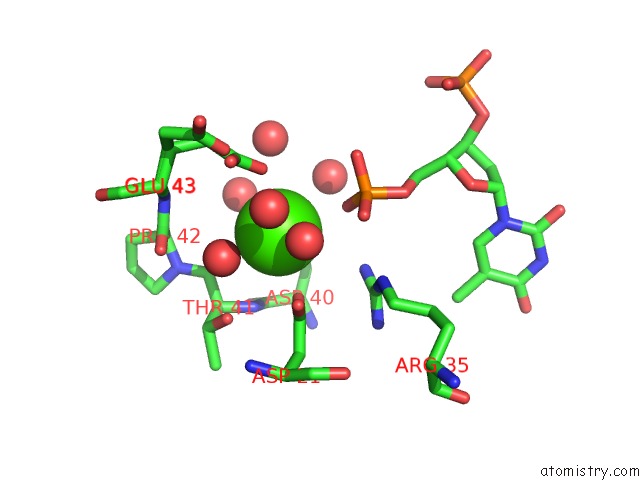
Mono view
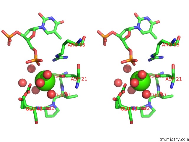
Stereo pair view
Mono view
Stereo pair view
A full contact list of Calcium with other atoms in the Ca binding
site number 1 of Crystal Structure of Staphylococcal Nuclease Variant Delta+Phs I92A/L25A at Cryogenic Temperature within 5.0Å range:
|
Reference:
J.A.Caro,
J.L.Schlessman,
B.Garcia-Moreno E..
Pressure Effects in Proteins To Be Published.
Page generated: Tue Jul 8 19:55:38 2025
Last articles
Mg in 6HW1Mg in 6HVX
Mg in 6HQB
Mg in 6HVW
Mg in 6HVV
Mg in 6HVU
Mg in 6HVT
Mg in 6HVS
Mg in 6HVR
Mg in 6HVA