Calcium »
PDB 4fsd-4gg1 »
4g26 »
Calcium in PDB 4g26: Crystal Structure of Proteinaceous Rnase P 1 (PRORP1) From A. Thaliana with Ca
Enzymatic activity of Crystal Structure of Proteinaceous Rnase P 1 (PRORP1) From A. Thaliana with Ca
All present enzymatic activity of Crystal Structure of Proteinaceous Rnase P 1 (PRORP1) From A. Thaliana with Ca:
3.1.26.5;
3.1.26.5;
Protein crystallography data
The structure of Crystal Structure of Proteinaceous Rnase P 1 (PRORP1) From A. Thaliana with Ca, PDB code: 4g26
was solved by
M.Koutmos,
M.J.Howard,
C.A.Fierke,
with X-Ray Crystallography technique. A brief refinement statistics is given in the table below:
| Resolution Low / High (Å) | 43.09 / 1.75 |
| Space group | P 21 21 21 |
| Cell size a, b, c (Å), α, β, γ (°) | 41.790, 111.883, 140.073, 90.00, 90.00, 90.00 |
| R / Rfree (%) | 16.2 / 20.9 |
Other elements in 4g26:
The structure of Crystal Structure of Proteinaceous Rnase P 1 (PRORP1) From A. Thaliana with Ca also contains other interesting chemical elements:
| Zinc | (Zn) | 1 atom |
Calcium Binding Sites:
The binding sites of Calcium atom in the Crystal Structure of Proteinaceous Rnase P 1 (PRORP1) From A. Thaliana with Ca
(pdb code 4g26). This binding sites where shown within
5.0 Angstroms radius around Calcium atom.
In total 5 binding sites of Calcium where determined in the Crystal Structure of Proteinaceous Rnase P 1 (PRORP1) From A. Thaliana with Ca, PDB code: 4g26:
Jump to Calcium binding site number: 1; 2; 3; 4; 5;
In total 5 binding sites of Calcium where determined in the Crystal Structure of Proteinaceous Rnase P 1 (PRORP1) From A. Thaliana with Ca, PDB code: 4g26:
Jump to Calcium binding site number: 1; 2; 3; 4; 5;
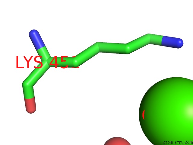
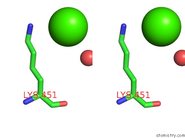
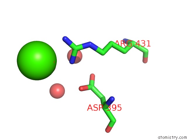
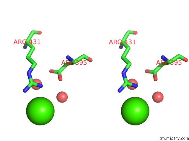
Calcium binding site 1 out of 5 in 4g26
Go back to
Calcium binding site 1 out
of 5 in the Crystal Structure of Proteinaceous Rnase P 1 (PRORP1) From A. Thaliana with Ca
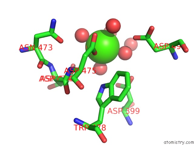
Mono view
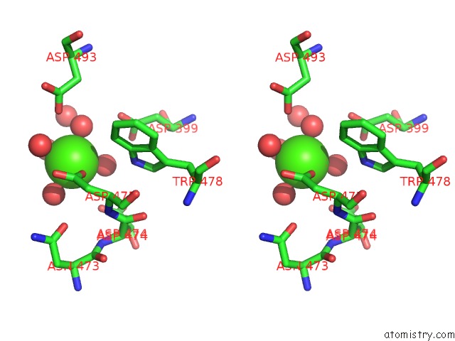
Stereo pair view
Mono view
Stereo pair view
A full contact list of Calcium with other atoms in the Ca binding
site number 1 of Crystal Structure of Proteinaceous Rnase P 1 (PRORP1) From A. Thaliana with Ca within 5.0Å range:
|
Calcium binding site 2 out of 5 in 4g26
Go back to
Calcium binding site 2 out
of 5 in the Crystal Structure of Proteinaceous Rnase P 1 (PRORP1) From A. Thaliana with Ca
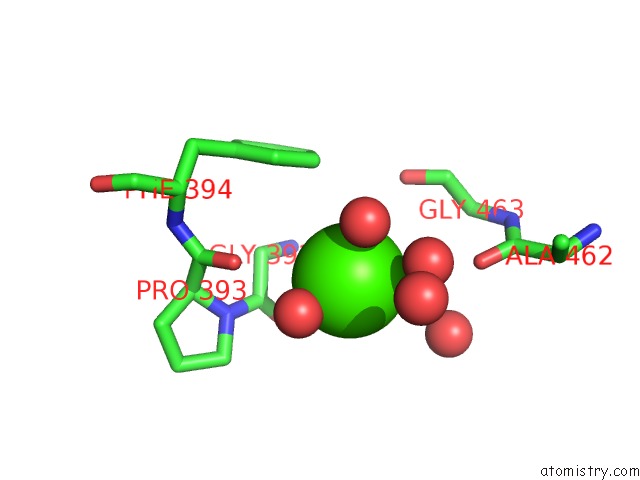
Mono view
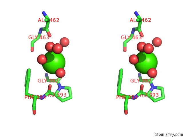
Stereo pair view
Mono view
Stereo pair view
A full contact list of Calcium with other atoms in the Ca binding
site number 2 of Crystal Structure of Proteinaceous Rnase P 1 (PRORP1) From A. Thaliana with Ca within 5.0Å range:
|
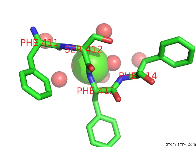
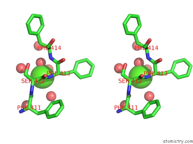
Calcium binding site 3 out of 5 in 4g26
Go back to
Calcium binding site 3 out
of 5 in the Crystal Structure of Proteinaceous Rnase P 1 (PRORP1) From A. Thaliana with Ca
Mono view
Stereo pair view
Mono view
Stereo pair view
A full contact list of Calcium with other atoms in the Ca binding
site number 3 of Crystal Structure of Proteinaceous Rnase P 1 (PRORP1) From A. Thaliana with Ca within 5.0Å range:
|
Calcium binding site 4 out of 5 in 4g26
Go back to
Calcium binding site 4 out
of 5 in the Crystal Structure of Proteinaceous Rnase P 1 (PRORP1) From A. Thaliana with Ca
Mono view
Stereo pair view
Mono view
Stereo pair view
A full contact list of Calcium with other atoms in the Ca binding
site number 4 of Crystal Structure of Proteinaceous Rnase P 1 (PRORP1) From A. Thaliana with Ca within 5.0Å range:
|
Calcium binding site 5 out of 5 in 4g26
Go back to
Calcium binding site 5 out
of 5 in the Crystal Structure of Proteinaceous Rnase P 1 (PRORP1) From A. Thaliana with Ca
Mono view
Stereo pair view
Mono view
Stereo pair view
A full contact list of Calcium with other atoms in the Ca binding
site number 5 of Crystal Structure of Proteinaceous Rnase P 1 (PRORP1) From A. Thaliana with Ca within 5.0Å range:
|
Reference:
M.J.Howard,
W.H.Lim,
C.A.Fierke,
M.Koutmos.
Mitochondrial Ribonuclease P Structure Provides Insight Into the Evolution of Catalytic Strategies For Precursor-Trna 5' Processing. Proc.Natl.Acad.Sci.Usa V. 109 16149 2012.
ISSN: ISSN 0027-8424
PubMed: 22991464
DOI: 10.1073/PNAS.1209062109
Page generated: Tue Jul 8 20:12:04 2025
ISSN: ISSN 0027-8424
PubMed: 22991464
DOI: 10.1073/PNAS.1209062109
Last articles
Mg in 4PJNMg in 4PJL
Mg in 4PJK
Mg in 4PJM
Mg in 4PJ0
Mg in 4PBU
Mg in 4PJ1
Mg in 4PJJ
Mg in 4PJ3
Mg in 4PJ2