Calcium »
PDB 4gq7-4gzs »
4gx0 »
Calcium in PDB 4gx0: Crystal Structure of the Gsuk L97D Mutant
Protein crystallography data
The structure of Crystal Structure of the Gsuk L97D Mutant, PDB code: 4gx0
was solved by
C.Kong,
W.Zeng,
S.Ye,
L.Chen,
D.B.Sauer,
Y.Lam,
M.G.Derebe,
Y.Jiang,
with X-Ray Crystallography technique. A brief refinement statistics is given in the table below:
| Resolution Low / High (Å) | 30.94 / 2.60 |
| Space group | C 1 2 1 |
| Cell size a, b, c (Å), α, β, γ (°) | 232.934, 111.670, 164.133, 90.00, 134.47, 90.00 |
| R / Rfree (%) | 20.3 / 24.9 |
Other elements in 4gx0:
The structure of Crystal Structure of the Gsuk L97D Mutant also contains other interesting chemical elements:
| Potassium | (K) | 12 atoms |
| Zinc | (Zn) | 4 atoms |
Calcium Binding Sites:
The binding sites of Calcium atom in the Crystal Structure of the Gsuk L97D Mutant
(pdb code 4gx0). This binding sites where shown within
5.0 Angstroms radius around Calcium atom.
In total 4 binding sites of Calcium where determined in the Crystal Structure of the Gsuk L97D Mutant, PDB code: 4gx0:
Jump to Calcium binding site number: 1; 2; 3; 4;
In total 4 binding sites of Calcium where determined in the Crystal Structure of the Gsuk L97D Mutant, PDB code: 4gx0:
Jump to Calcium binding site number: 1; 2; 3; 4;
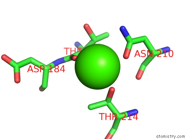
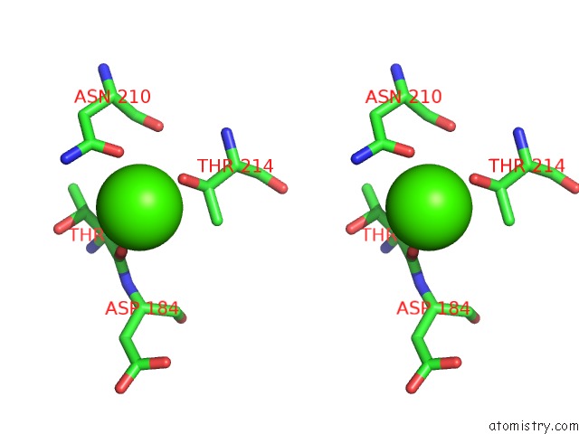
Calcium binding site 1 out of 4 in 4gx0
Go back to
Calcium binding site 1 out
of 4 in the Crystal Structure of the Gsuk L97D Mutant
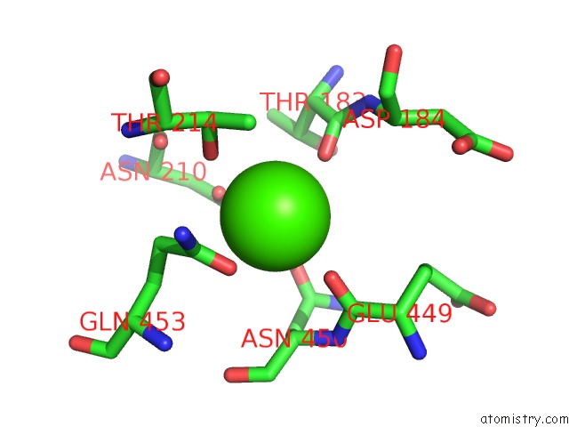
Mono view
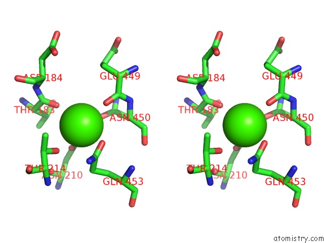
Stereo pair view
Mono view
Stereo pair view
A full contact list of Calcium with other atoms in the Ca binding
site number 1 of Crystal Structure of the Gsuk L97D Mutant within 5.0Å range:
|
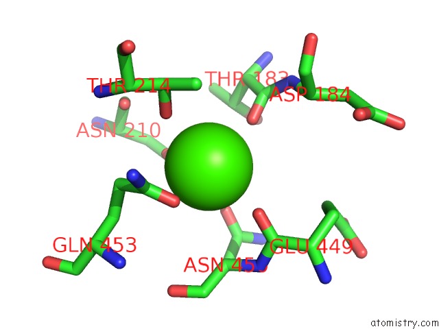
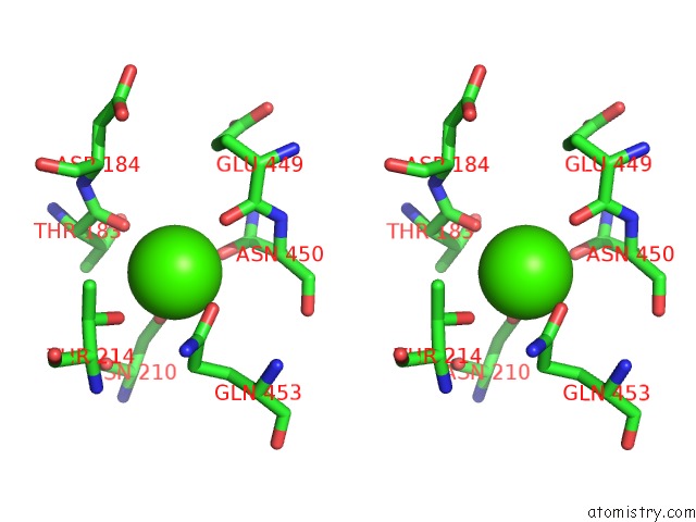
Calcium binding site 2 out of 4 in 4gx0
Go back to
Calcium binding site 2 out
of 4 in the Crystal Structure of the Gsuk L97D Mutant
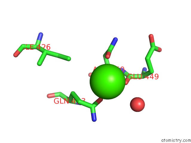
Mono view
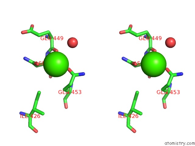
Stereo pair view
Mono view
Stereo pair view
A full contact list of Calcium with other atoms in the Ca binding
site number 2 of Crystal Structure of the Gsuk L97D Mutant within 5.0Å range:
|
Calcium binding site 3 out of 4 in 4gx0
Go back to
Calcium binding site 3 out
of 4 in the Crystal Structure of the Gsuk L97D Mutant
Mono view
Stereo pair view
Mono view
Stereo pair view
A full contact list of Calcium with other atoms in the Ca binding
site number 3 of Crystal Structure of the Gsuk L97D Mutant within 5.0Å range:
|
Calcium binding site 4 out of 4 in 4gx0
Go back to
Calcium binding site 4 out
of 4 in the Crystal Structure of the Gsuk L97D Mutant
Mono view
Stereo pair view
Mono view
Stereo pair view
A full contact list of Calcium with other atoms in the Ca binding
site number 4 of Crystal Structure of the Gsuk L97D Mutant within 5.0Å range:
|
Reference:
C.Kong,
W.Zeng,
S.Ye,
L.Chen,
D.B.Sauer,
Y.Lam,
M.G.Derebe,
Y.Jiang.
Distinct Gating Mechanisms Revealed By the Structures of A Multi-Ligand Gated K(+) Channel. Elife V. 1 00184 2012.
PubMed: 23240087
DOI: 10.7554/ELIFE.00184
Page generated: Tue Jul 8 22:26:45 2025
PubMed: 23240087
DOI: 10.7554/ELIFE.00184
Last articles
Mg in 4W5OMg in 4W5J
Mg in 4W5N
Mg in 4V2I
Mg in 4V3R
Mg in 4V26
Mg in 4V2G
Mg in 4V1T
Mg in 4V25
Mg in 4V1V