Calcium »
PDB 4gq7-4gzs »
4gy2 »
Calcium in PDB 4gy2: Crystal Structure of Apo-Ia-Actin Complex
Enzymatic activity of Crystal Structure of Apo-Ia-Actin Complex
All present enzymatic activity of Crystal Structure of Apo-Ia-Actin Complex:
2.4.2.31;
2.4.2.31;
Protein crystallography data
The structure of Crystal Structure of Apo-Ia-Actin Complex, PDB code: 4gy2
was solved by
T.Tsurumura,
M.Oda,
M.Nagahama,
H.Tsuge,
with X-Ray Crystallography technique. A brief refinement statistics is given in the table below:
| Resolution Low / High (Å) | 37.19 / 2.71 |
| Space group | P 21 21 21 |
| Cell size a, b, c (Å), α, β, γ (°) | 54.442, 135.872, 153.946, 90.00, 90.00, 90.00 |
| R / Rfree (%) | 19.9 / 25.7 |
Calcium Binding Sites:
The binding sites of Calcium atom in the Crystal Structure of Apo-Ia-Actin Complex
(pdb code 4gy2). This binding sites where shown within
5.0 Angstroms radius around Calcium atom.
In total only one binding site of Calcium was determined in the Crystal Structure of Apo-Ia-Actin Complex, PDB code: 4gy2:
In total only one binding site of Calcium was determined in the Crystal Structure of Apo-Ia-Actin Complex, PDB code: 4gy2:
Calcium binding site 1 out of 1 in 4gy2
Go back to
Calcium binding site 1 out
of 1 in the Crystal Structure of Apo-Ia-Actin Complex
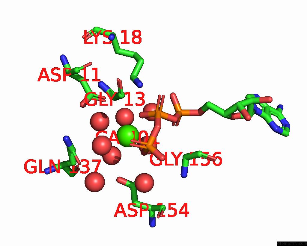
Mono view
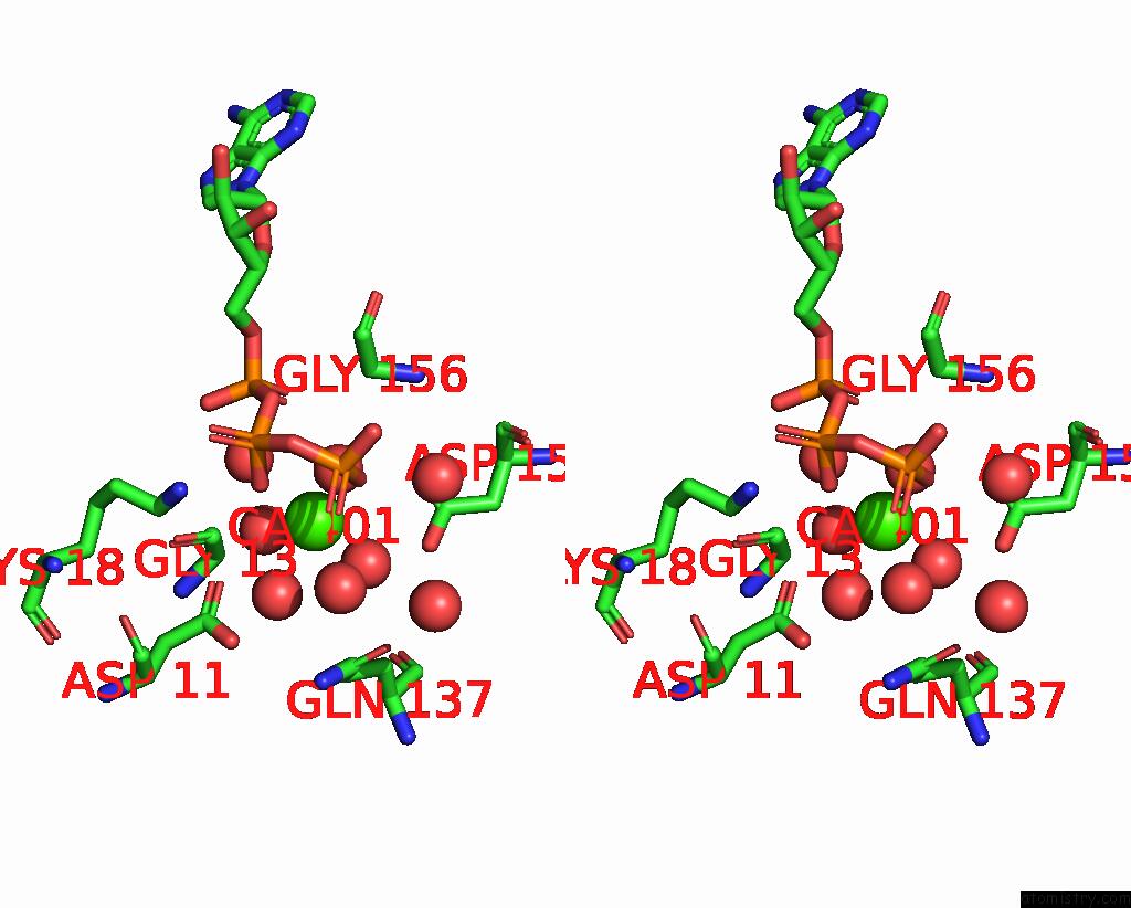
Stereo pair view
Mono view
Stereo pair view
A full contact list of Calcium with other atoms in the Ca binding
site number 1 of Crystal Structure of Apo-Ia-Actin Complex within 5.0Å range:
|
Reference:
T.Tsurumura,
Y.Tsumori,
H.Qiu,
M.Oda,
J.Sakurai,
M.Nagahama,
H.Tsuge.
Arginine Adp-Ribosylation Mechanism Based on Structural Snapshots of Iota-Toxin and Actin Complex Proc.Natl.Acad.Sci.Usa V. 110 4267 2013.
ISSN: ISSN 0027-8424
PubMed: 23382240
DOI: 10.1073/PNAS.1217227110
Page generated: Tue Jul 8 22:27:41 2025
ISSN: ISSN 0027-8424
PubMed: 23382240
DOI: 10.1073/PNAS.1217227110
Last articles
Mg in 4W5OMg in 4W5J
Mg in 4W5N
Mg in 4V2I
Mg in 4V3R
Mg in 4V26
Mg in 4V2G
Mg in 4V1T
Mg in 4V25
Mg in 4V1V