Calcium »
PDB 4h84-4hte »
4hjf »
Calcium in PDB 4hjf: Eal Domain of Phosphodiesterase Pdea in Complex with C-Di-Gmp and Ca++
Protein crystallography data
The structure of Eal Domain of Phosphodiesterase Pdea in Complex with C-Di-Gmp and Ca++, PDB code: 4hjf
was solved by
E.V.Filippova,
G.Minasov,
L.Shuvalova,
O.Kiryukhina,
C.Massa,
T.Schirmer,
A.Joachimiak,
W.F.Anderson,
Midwest Center For Structural Genomics(Mcsg),
with X-Ray Crystallography technique. A brief refinement statistics is given in the table below:
| Resolution Low / High (Å) | 29.22 / 1.75 |
| Space group | P 21 21 21 |
| Cell size a, b, c (Å), α, β, γ (°) | 62.202, 63.186, 66.190, 90.00, 90.00, 90.00 |
| R / Rfree (%) | 16.8 / 21.4 |
Calcium Binding Sites:
The binding sites of Calcium atom in the Eal Domain of Phosphodiesterase Pdea in Complex with C-Di-Gmp and Ca++
(pdb code 4hjf). This binding sites where shown within
5.0 Angstroms radius around Calcium atom.
In total 3 binding sites of Calcium where determined in the Eal Domain of Phosphodiesterase Pdea in Complex with C-Di-Gmp and Ca++, PDB code: 4hjf:
Jump to Calcium binding site number: 1; 2; 3;
In total 3 binding sites of Calcium where determined in the Eal Domain of Phosphodiesterase Pdea in Complex with C-Di-Gmp and Ca++, PDB code: 4hjf:
Jump to Calcium binding site number: 1; 2; 3;
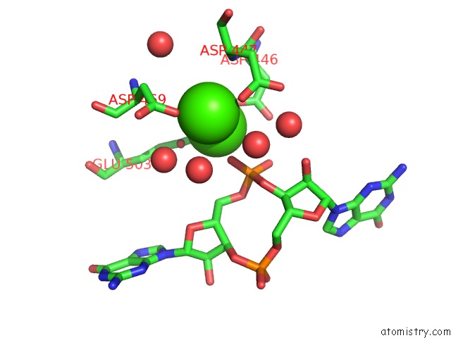
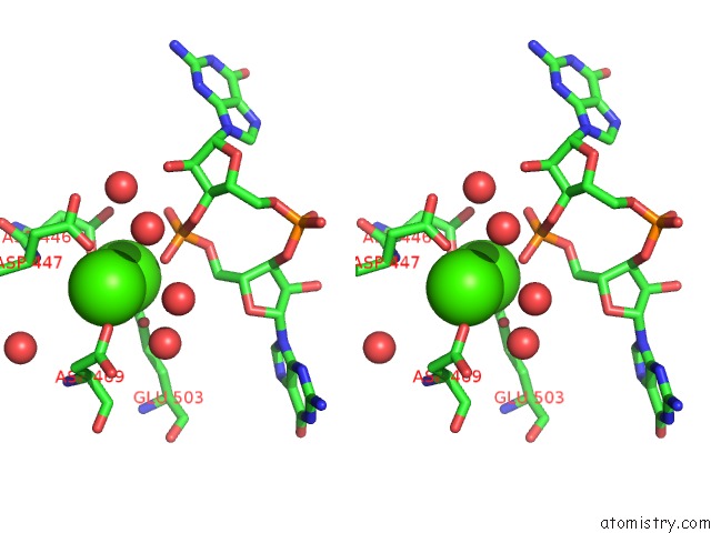
Calcium binding site 1 out of 3 in 4hjf
Go back to
Calcium binding site 1 out
of 3 in the Eal Domain of Phosphodiesterase Pdea in Complex with C-Di-Gmp and Ca++
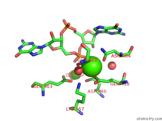
Mono view
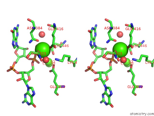
Stereo pair view
Mono view
Stereo pair view
A full contact list of Calcium with other atoms in the Ca binding
site number 1 of Eal Domain of Phosphodiesterase Pdea in Complex with C-Di-Gmp and Ca++ within 5.0Å range:
|
Calcium binding site 2 out of 3 in 4hjf
Go back to
Calcium binding site 2 out
of 3 in the Eal Domain of Phosphodiesterase Pdea in Complex with C-Di-Gmp and Ca++
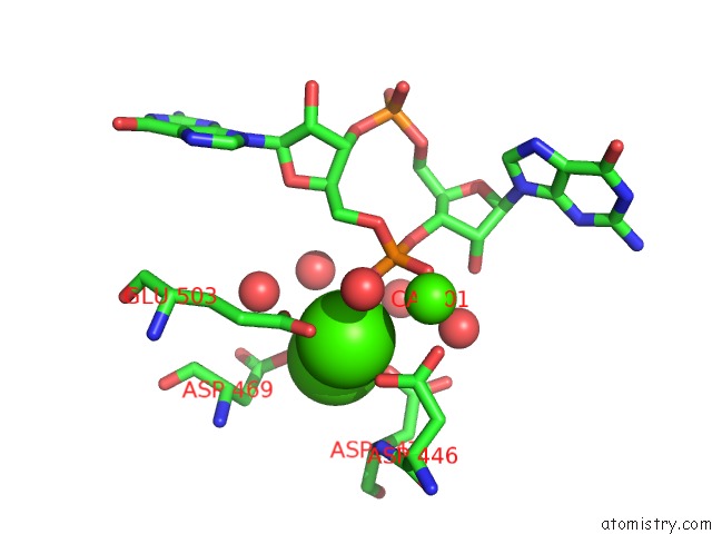
Mono view
Stereo pair view
Mono view
Stereo pair view
A full contact list of Calcium with other atoms in the Ca binding
site number 2 of Eal Domain of Phosphodiesterase Pdea in Complex with C-Di-Gmp and Ca++ within 5.0Å range:
|
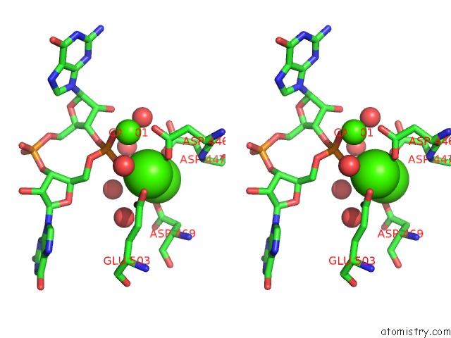
Calcium binding site 3 out of 3 in 4hjf
Go back to
Calcium binding site 3 out
of 3 in the Eal Domain of Phosphodiesterase Pdea in Complex with C-Di-Gmp and Ca++
Mono view
Stereo pair view
Mono view
Stereo pair view
A full contact list of Calcium with other atoms in the Ca binding
site number 3 of Eal Domain of Phosphodiesterase Pdea in Complex with C-Di-Gmp and Ca++ within 5.0Å range:
|
Reference:
E.V.Filippova,
G.Minasov,
L.Shuvalova,
O.Kiryukhina,
C.Massa,
T.Schirmer,
A.Joachimiak,
W.F.Anderson.
Crystal Structure of Eal Domain From Caulobacter Crescentus in Complex with C-Di-Gmp and Ca To Be Published.
Page generated: Tue Jul 8 22:40:53 2025
Last articles
Mg in 5T4YMg in 5T63
Mg in 5T5I
Mg in 5T5C
Mg in 5T3R
Mg in 5T40
Mg in 5T41
Mg in 5T45
Mg in 5T3K
Mg in 5T14