Calcium »
PDB 4hvk-4i9x »
4hxb »
Calcium in PDB 4hxb: Crystal Structure of 6B9 Fab
Protein crystallography data
The structure of Crystal Structure of 6B9 Fab, PDB code: 4hxb
was solved by
A.R.Johal,
H.C.Jarrell,
N.H.Khieu,
J.A.Letts,
R.C.Landry,
W.Jachymek,
Q.Yang,
H.J.Jennings,
J.R.Brisson,
S.V.Evans,
with X-Ray Crystallography technique. A brief refinement statistics is given in the table below:
| Resolution Low / High (Å) | 20.00 / 2.45 |
| Space group | P 21 21 2 |
| Cell size a, b, c (Å), α, β, γ (°) | 89.641, 131.990, 36.921, 90.00, 90.00, 90.00 |
| R / Rfree (%) | 19.7 / 27.9 |
Calcium Binding Sites:
The binding sites of Calcium atom in the Crystal Structure of 6B9 Fab
(pdb code 4hxb). This binding sites where shown within
5.0 Angstroms radius around Calcium atom.
In total 2 binding sites of Calcium where determined in the Crystal Structure of 6B9 Fab, PDB code: 4hxb:
Jump to Calcium binding site number: 1; 2;
In total 2 binding sites of Calcium where determined in the Crystal Structure of 6B9 Fab, PDB code: 4hxb:
Jump to Calcium binding site number: 1; 2;
Calcium binding site 1 out of 2 in 4hxb
Go back to
Calcium binding site 1 out
of 2 in the Crystal Structure of 6B9 Fab
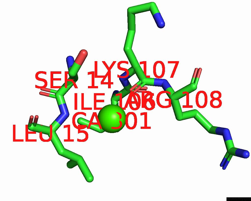
Mono view

Stereo pair view
Mono view
Stereo pair view
A full contact list of Calcium with other atoms in the Ca binding
site number 1 of Crystal Structure of 6B9 Fab within 5.0Å range:
|
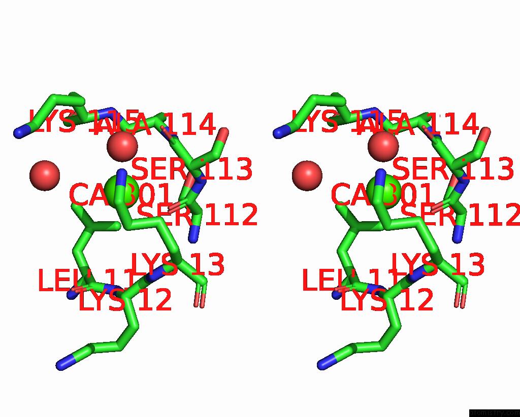
Calcium binding site 2 out of 2 in 4hxb
Go back to
Calcium binding site 2 out
of 2 in the Crystal Structure of 6B9 Fab

Mono view
Stereo pair view
Mono view
Stereo pair view
A full contact list of Calcium with other atoms in the Ca binding
site number 2 of Crystal Structure of 6B9 Fab within 5.0Å range:
|
Reference:
A.R.Johal,
H.C.Jarrell,
J.A.Letts,
N.H.Khieu,
R.C.Landry,
W.Jachymek,
Q.Yang,
H.J.Jennings,
J.R.Brisson,
S.V.Evans.
The Antigen-Binding Site of An N-Propionylated Polysialic Acid-Specific Antibody Protective Against Group B Meningococci Is Consistent with Extended Epitopes. Glycobiology V. 23 946 2013.
ISSN: ISSN 0959-6658
PubMed: 23704298
DOI: 10.1093/GLYCOB/CWT031
Page generated: Tue Jul 8 22:45:15 2025
ISSN: ISSN 0959-6658
PubMed: 23704298
DOI: 10.1093/GLYCOB/CWT031
Last articles
Mg in 6TRAMg in 6TR4
Mg in 6TR3
Mg in 6TMF
Mg in 6TQO
Mg in 6TQN
Mg in 6TQF
Mg in 6TQE
Mg in 6TQB
Mg in 6TQA