Calcium »
PDB 4iai-4iun »
4ild »
Calcium in PDB 4ild: Crystal Structure of Truncated Bovine Viral Diarrhea Virus 1 E2 Envelope Protein
Protein crystallography data
The structure of Crystal Structure of Truncated Bovine Viral Diarrhea Virus 1 E2 Envelope Protein, PDB code: 4ild
was solved by
Y.Modis,
Y.Li,
J.Wang,
with X-Ray Crystallography technique. A brief refinement statistics is given in the table below:
| Resolution Low / High (Å) | 47.97 / 3.27 |
| Space group | C 1 2 1 |
| Cell size a, b, c (Å), α, β, γ (°) | 136.720, 54.450, 95.920, 90.00, 92.23, 90.00 |
| R / Rfree (%) | 24.6 / 28.9 |
Other elements in 4ild:
The structure of Crystal Structure of Truncated Bovine Viral Diarrhea Virus 1 E2 Envelope Protein also contains other interesting chemical elements:
| Uranium | (U) | 4 atoms |
Calcium Binding Sites:
The binding sites of Calcium atom in the Crystal Structure of Truncated Bovine Viral Diarrhea Virus 1 E2 Envelope Protein
(pdb code 4ild). This binding sites where shown within
5.0 Angstroms radius around Calcium atom.
In total 4 binding sites of Calcium where determined in the Crystal Structure of Truncated Bovine Viral Diarrhea Virus 1 E2 Envelope Protein, PDB code: 4ild:
Jump to Calcium binding site number: 1; 2; 3; 4;
In total 4 binding sites of Calcium where determined in the Crystal Structure of Truncated Bovine Viral Diarrhea Virus 1 E2 Envelope Protein, PDB code: 4ild:
Jump to Calcium binding site number: 1; 2; 3; 4;
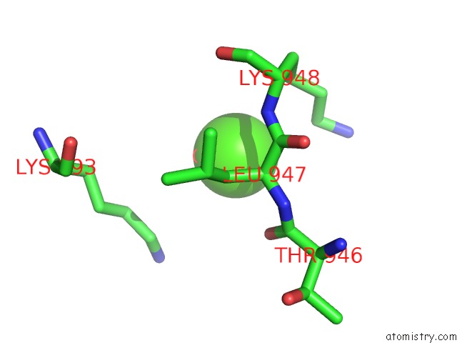
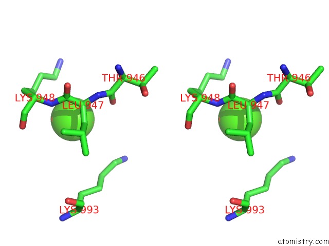
Calcium binding site 1 out of 4 in 4ild
Go back to
Calcium binding site 1 out
of 4 in the Crystal Structure of Truncated Bovine Viral Diarrhea Virus 1 E2 Envelope Protein
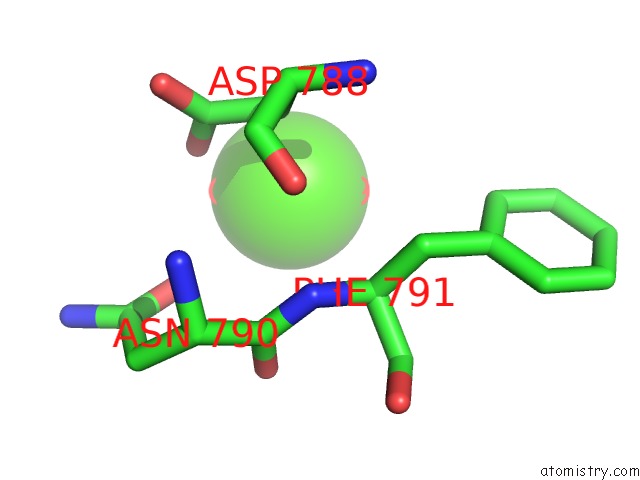
Mono view
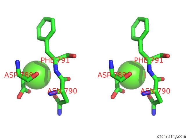
Stereo pair view
Mono view
Stereo pair view
A full contact list of Calcium with other atoms in the Ca binding
site number 1 of Crystal Structure of Truncated Bovine Viral Diarrhea Virus 1 E2 Envelope Protein within 5.0Å range:
|
Calcium binding site 2 out of 4 in 4ild
Go back to
Calcium binding site 2 out
of 4 in the Crystal Structure of Truncated Bovine Viral Diarrhea Virus 1 E2 Envelope Protein
Mono view
Stereo pair view
Mono view
Stereo pair view
A full contact list of Calcium with other atoms in the Ca binding
site number 2 of Crystal Structure of Truncated Bovine Viral Diarrhea Virus 1 E2 Envelope Protein within 5.0Å range:
|
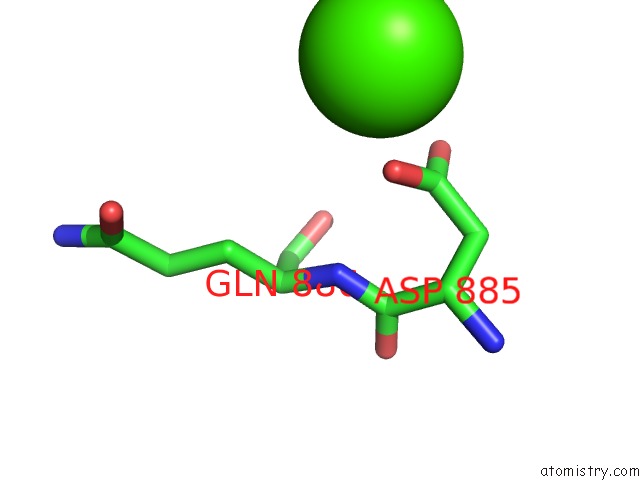
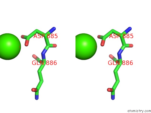
Calcium binding site 3 out of 4 in 4ild
Go back to
Calcium binding site 3 out
of 4 in the Crystal Structure of Truncated Bovine Viral Diarrhea Virus 1 E2 Envelope Protein
Mono view
Stereo pair view
Mono view
Stereo pair view
A full contact list of Calcium with other atoms in the Ca binding
site number 3 of Crystal Structure of Truncated Bovine Viral Diarrhea Virus 1 E2 Envelope Protein within 5.0Å range:
|
Calcium binding site 4 out of 4 in 4ild
Go back to
Calcium binding site 4 out
of 4 in the Crystal Structure of Truncated Bovine Viral Diarrhea Virus 1 E2 Envelope Protein
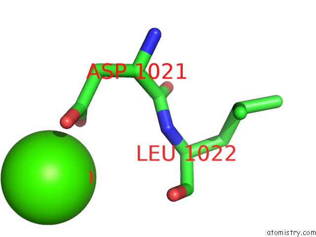
Mono view
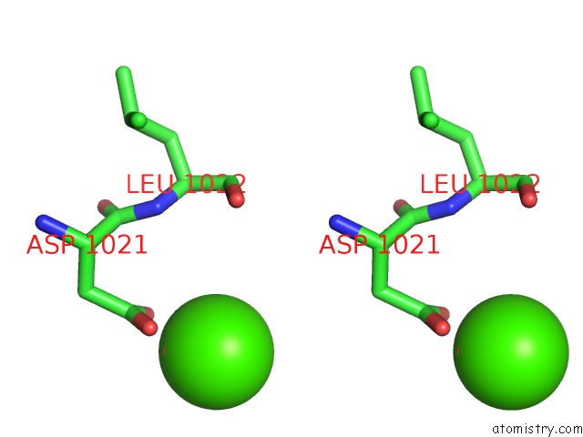
Stereo pair view
Mono view
Stereo pair view
A full contact list of Calcium with other atoms in the Ca binding
site number 4 of Crystal Structure of Truncated Bovine Viral Diarrhea Virus 1 E2 Envelope Protein within 5.0Å range:
|
Reference:
Y.Li,
J.Wang,
R.Kanai,
Y.Modis.
Crystal Structure of Glycoprotein E2 From Bovine Viral Diarrhea Virus. Proc.Natl.Acad.Sci.Usa V. 110 6805 2013.
ISSN: ISSN 0027-8424
PubMed: 23569276
DOI: 10.1073/PNAS.1300524110
Page generated: Tue Jul 8 22:59:15 2025
ISSN: ISSN 0027-8424
PubMed: 23569276
DOI: 10.1073/PNAS.1300524110
Last articles
Mg in 5SDZMg in 5SE1
Mg in 5SDX
Mg in 5SDT
Mg in 5SDY
Mg in 5SDW
Mg in 5SDV
Mg in 5SDU
Mg in 5SCL
Mg in 5SCI