Calcium »
PDB 4l1i-4lls »
4lcv »
Calcium in PDB 4lcv: Crystal Structure of DOC2B C2A Domain
Protein crystallography data
The structure of Crystal Structure of DOC2B C2A Domain, PDB code: 4lcv
was solved by
M.Giladi,
L.Almagor,
J.A.Hirsch,
with X-Ray Crystallography technique. A brief refinement statistics is given in the table below:
| Resolution Low / High (Å) | 47.58 / 2.00 |
| Space group | P 1 21 1 |
| Cell size a, b, c (Å), α, β, γ (°) | 43.632, 95.151, 67.816, 90.00, 99.95, 90.00 |
| R / Rfree (%) | 18.7 / 23.2 |
Calcium Binding Sites:
The binding sites of Calcium atom in the Crystal Structure of DOC2B C2A Domain
(pdb code 4lcv). This binding sites where shown within
5.0 Angstroms radius around Calcium atom.
In total 2 binding sites of Calcium where determined in the Crystal Structure of DOC2B C2A Domain, PDB code: 4lcv:
Jump to Calcium binding site number: 1; 2;
In total 2 binding sites of Calcium where determined in the Crystal Structure of DOC2B C2A Domain, PDB code: 4lcv:
Jump to Calcium binding site number: 1; 2;
Calcium binding site 1 out of 2 in 4lcv
Go back to
Calcium binding site 1 out
of 2 in the Crystal Structure of DOC2B C2A Domain
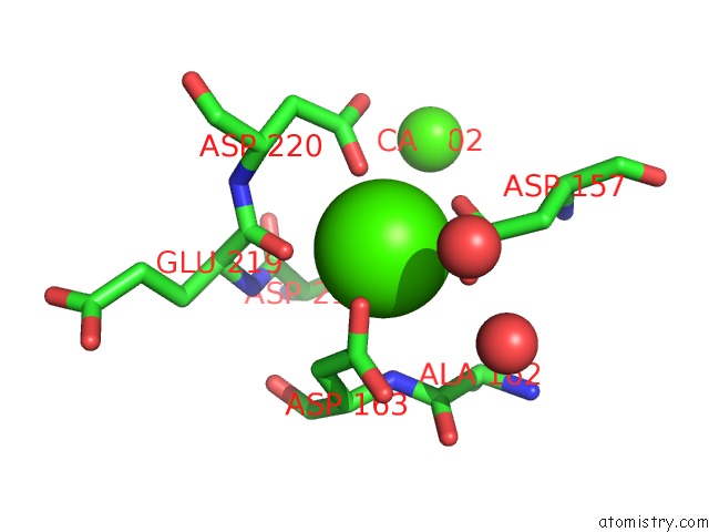
Mono view
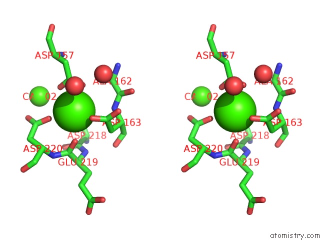
Stereo pair view
Mono view
Stereo pair view
A full contact list of Calcium with other atoms in the Ca binding
site number 1 of Crystal Structure of DOC2B C2A Domain within 5.0Å range:
|
Calcium binding site 2 out of 2 in 4lcv
Go back to
Calcium binding site 2 out
of 2 in the Crystal Structure of DOC2B C2A Domain
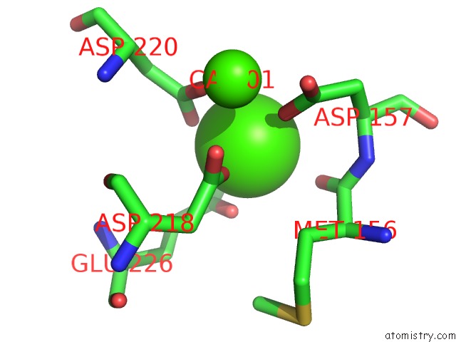
Mono view
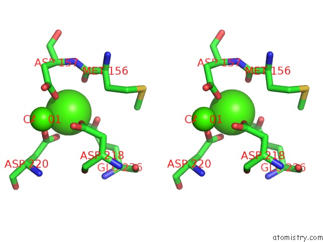
Stereo pair view
Mono view
Stereo pair view
A full contact list of Calcium with other atoms in the Ca binding
site number 2 of Crystal Structure of DOC2B C2A Domain within 5.0Å range:
|
Reference:
M.Giladi,
L.Michaeli,
L.Almagor,
D.Bar-On,
T.Buki,
U.Ashery,
D.Khananshvili,
J.A.Hirsch.
The C2B Domain Is the Primary Ca(2+) Sensor in DOC2B: A Structural and Functional Analysis. J.Mol.Biol. V. 425 4629 2013.
ISSN: ISSN 0022-2836
PubMed: 23994332
DOI: 10.1016/J.JMB.2013.08.017
Page generated: Tue Jul 8 23:48:08 2025
ISSN: ISSN 0022-2836
PubMed: 23994332
DOI: 10.1016/J.JMB.2013.08.017
Last articles
I in 3WN5I in 3WYX
I in 3WGW
I in 3WD6
I in 3WB5
I in 3W31
I in 3WB4
I in 3W1N
I in 3W0F
I in 3W2Z