Calcium »
PDB 4m18-4m8u »
4m6y »
Calcium in PDB 4m6y: Mutant Structure of Methyltransferase From Streptomyces Hygroscopicus Complexed with S-Adenosyl-L-Homocysteine and Methylphenylpyruvic Acid
Protein crystallography data
The structure of Mutant Structure of Methyltransferase From Streptomyces Hygroscopicus Complexed with S-Adenosyl-L-Homocysteine and Methylphenylpyruvic Acid, PDB code: 4m6y
was solved by
Y.C.Liu,
X.W.Zou,
H.C.Chan,
C.J.Huang,
T.L.Li,
with X-Ray Crystallography technique. A brief refinement statistics is given in the table below:
| Resolution Low / High (Å) | 29.34 / 2.50 |
| Space group | P 21 21 21 |
| Cell size a, b, c (Å), α, β, γ (°) | 57.160, 89.626, 136.622, 90.00, 90.00, 90.00 |
| R / Rfree (%) | 17.8 / 25.5 |
Other elements in 4m6y:
The structure of Mutant Structure of Methyltransferase From Streptomyces Hygroscopicus Complexed with S-Adenosyl-L-Homocysteine and Methylphenylpyruvic Acid also contains other interesting chemical elements:
| Iron | (Fe) | 2 atoms |
Calcium Binding Sites:
The binding sites of Calcium atom in the Mutant Structure of Methyltransferase From Streptomyces Hygroscopicus Complexed with S-Adenosyl-L-Homocysteine and Methylphenylpyruvic Acid
(pdb code 4m6y). This binding sites where shown within
5.0 Angstroms radius around Calcium atom.
In total 4 binding sites of Calcium where determined in the Mutant Structure of Methyltransferase From Streptomyces Hygroscopicus Complexed with S-Adenosyl-L-Homocysteine and Methylphenylpyruvic Acid, PDB code: 4m6y:
Jump to Calcium binding site number: 1; 2; 3; 4;
In total 4 binding sites of Calcium where determined in the Mutant Structure of Methyltransferase From Streptomyces Hygroscopicus Complexed with S-Adenosyl-L-Homocysteine and Methylphenylpyruvic Acid, PDB code: 4m6y:
Jump to Calcium binding site number: 1; 2; 3; 4;
Calcium binding site 1 out of 4 in 4m6y
Go back to
Calcium binding site 1 out
of 4 in the Mutant Structure of Methyltransferase From Streptomyces Hygroscopicus Complexed with S-Adenosyl-L-Homocysteine and Methylphenylpyruvic Acid
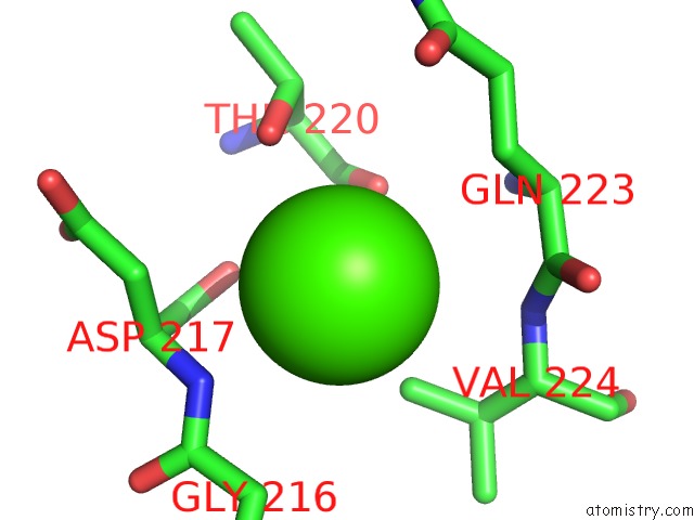
Mono view

Stereo pair view
Mono view
Stereo pair view
A full contact list of Calcium with other atoms in the Ca binding
site number 1 of Mutant Structure of Methyltransferase From Streptomyces Hygroscopicus Complexed with S-Adenosyl-L-Homocysteine and Methylphenylpyruvic Acid within 5.0Å range:
|
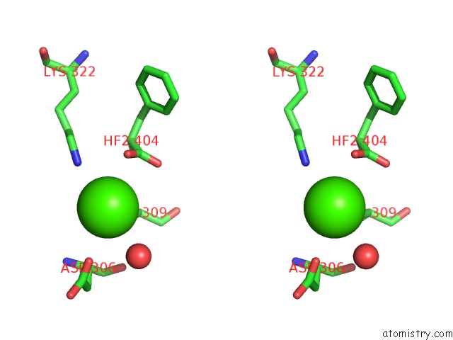
Calcium binding site 2 out of 4 in 4m6y
Go back to
Calcium binding site 2 out
of 4 in the Mutant Structure of Methyltransferase From Streptomyces Hygroscopicus Complexed with S-Adenosyl-L-Homocysteine and Methylphenylpyruvic Acid

Mono view

Stereo pair view
Mono view
Stereo pair view
A full contact list of Calcium with other atoms in the Ca binding
site number 2 of Mutant Structure of Methyltransferase From Streptomyces Hygroscopicus Complexed with S-Adenosyl-L-Homocysteine and Methylphenylpyruvic Acid within 5.0Å range:
|
Calcium binding site 3 out of 4 in 4m6y
Go back to
Calcium binding site 3 out
of 4 in the Mutant Structure of Methyltransferase From Streptomyces Hygroscopicus Complexed with S-Adenosyl-L-Homocysteine and Methylphenylpyruvic Acid

Mono view

Stereo pair view
Mono view
Stereo pair view
A full contact list of Calcium with other atoms in the Ca binding
site number 3 of Mutant Structure of Methyltransferase From Streptomyces Hygroscopicus Complexed with S-Adenosyl-L-Homocysteine and Methylphenylpyruvic Acid within 5.0Å range:
|
Calcium binding site 4 out of 4 in 4m6y
Go back to
Calcium binding site 4 out
of 4 in the Mutant Structure of Methyltransferase From Streptomyces Hygroscopicus Complexed with S-Adenosyl-L-Homocysteine and Methylphenylpyruvic Acid

Mono view
Stereo pair view
Mono view
Stereo pair view
A full contact list of Calcium with other atoms in the Ca binding
site number 4 of Mutant Structure of Methyltransferase From Streptomyces Hygroscopicus Complexed with S-Adenosyl-L-Homocysteine and Methylphenylpyruvic Acid within 5.0Å range:
|
Reference:
X.W.Zou,
Y.C.Liu,
N.S.Hsu,
C.J.Huang,
S.Y.Lyu,
H.C.Chan,
C.Y.Chang,
H.W.Yeh,
K.H.Lin,
C.J.Wu,
M.D.Tsai,
T.L.Li.
Structure and Mechanism of A Nonhaem-Iron Sam-Dependent C-Methyltransferase and Its Engineering to A Hydratase and An O-Methyltransferase Acta Crystallogr.,Sect.D V. 70 1549 2014.
ISSN: ISSN 0907-4449
PubMed: 24914966
DOI: 10.1107/S1399004714005239
Page generated: Wed Jul 9 00:15:38 2025
ISSN: ISSN 0907-4449
PubMed: 24914966
DOI: 10.1107/S1399004714005239
Last articles
W in 9FPPW in 8PRM
W in 9QM1
W in 9QM0
W in 9OJ3
W in 9MQX
W in 9FP4
W in 9BEO
W in 9BEM
W in 8P2U