Calcium »
PDB 4niv-4o4h »
4niw »
Calcium in PDB 4niw: Crystal Structure of Trypsiligase (K60E/N143H/Y151H/D189K Trypsin) Orthorhombic Form
Enzymatic activity of Crystal Structure of Trypsiligase (K60E/N143H/Y151H/D189K Trypsin) Orthorhombic Form
All present enzymatic activity of Crystal Structure of Trypsiligase (K60E/N143H/Y151H/D189K Trypsin) Orthorhombic Form:
3.4.21.4;
3.4.21.4;
Protein crystallography data
The structure of Crystal Structure of Trypsiligase (K60E/N143H/Y151H/D189K Trypsin) Orthorhombic Form, PDB code: 4niw
was solved by
M.Schoepfel,
C.Parthier,
M.T.Stubbs,
with X-Ray Crystallography technique. A brief refinement statistics is given in the table below:
| Resolution Low / High (Å) | 44.06 / 1.31 |
| Space group | P 21 21 21 |
| Cell size a, b, c (Å), α, β, γ (°) | 54.522, 58.607, 66.831, 90.00, 90.00, 90.00 |
| R / Rfree (%) | 12.8 / 15.5 |
Calcium Binding Sites:
The binding sites of Calcium atom in the Crystal Structure of Trypsiligase (K60E/N143H/Y151H/D189K Trypsin) Orthorhombic Form
(pdb code 4niw). This binding sites where shown within
5.0 Angstroms radius around Calcium atom.
In total only one binding site of Calcium was determined in the Crystal Structure of Trypsiligase (K60E/N143H/Y151H/D189K Trypsin) Orthorhombic Form, PDB code: 4niw:
In total only one binding site of Calcium was determined in the Crystal Structure of Trypsiligase (K60E/N143H/Y151H/D189K Trypsin) Orthorhombic Form, PDB code: 4niw:
Calcium binding site 1 out of 1 in 4niw
Go back to
Calcium binding site 1 out
of 1 in the Crystal Structure of Trypsiligase (K60E/N143H/Y151H/D189K Trypsin) Orthorhombic Form
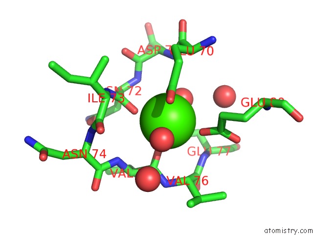
Mono view
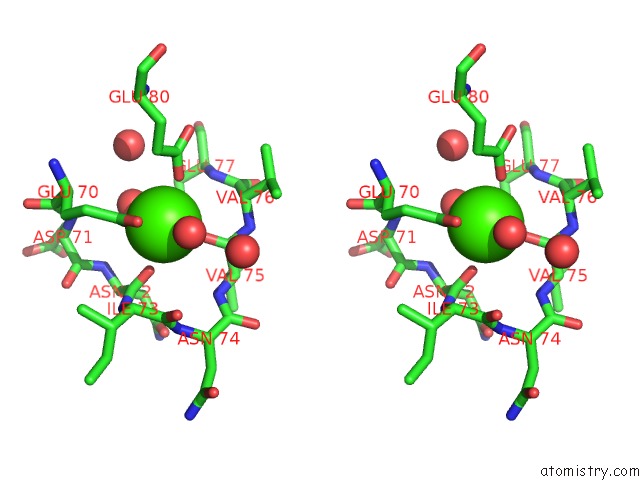
Stereo pair view
Mono view
Stereo pair view
A full contact list of Calcium with other atoms in the Ca binding
site number 1 of Crystal Structure of Trypsiligase (K60E/N143H/Y151H/D189K Trypsin) Orthorhombic Form within 5.0Å range:
|
Reference:
S.Liebscher,
M.Schopfel,
T.Aumuller,
A.Sharkhuukhen,
A.Pech,
E.Hoss,
C.Parthier,
G.Jahreis,
M.T.Stubbs,
F.Bordusa.
N-Terminal Protein Modification By Substrate-Activated Reverse Proteolysis. Angew.Chem.Int.Ed.Engl. V. 53 3024 2014.
ISSN: ISSN 1433-7851
PubMed: 24520050
DOI: 10.1002/ANIE.201307736
Page generated: Wed Jul 9 00:52:27 2025
ISSN: ISSN 1433-7851
PubMed: 24520050
DOI: 10.1002/ANIE.201307736
Last articles
Mg in 5XNAMg in 5XN9
Mg in 5XN2
Mg in 5XN1
Mg in 5XLZ
Mg in 5XLT
Mg in 5XM7
Mg in 5XM3
Mg in 5XLH
Mg in 5XKM