Calcium »
PDB 4opq-4p4f »
4p2y »
Calcium in PDB 4p2y: Crystal Structure of the Human Rage Ectodomain (Fragment VC1C2) in Complex with Mouse S100A6
Protein crystallography data
The structure of Crystal Structure of the Human Rage Ectodomain (Fragment VC1C2) in Complex with Mouse S100A6, PDB code: 4p2y
was solved by
L.Yatime,
G.R.Andersen,
with X-Ray Crystallography technique. A brief refinement statistics is given in the table below:
| Resolution Low / High (Å) | 27.24 / 2.30 |
| Space group | I 2 2 2 |
| Cell size a, b, c (Å), α, β, γ (°) | 76.780, 113.390, 140.320, 90.00, 90.00, 90.00 |
| R / Rfree (%) | 18.7 / 20.7 |
Other elements in 4p2y:
The structure of Crystal Structure of the Human Rage Ectodomain (Fragment VC1C2) in Complex with Mouse S100A6 also contains other interesting chemical elements:
| Chlorine | (Cl) | 6 atoms |
| Zinc | (Zn) | 6 atoms |
Calcium Binding Sites:
The binding sites of Calcium atom in the Crystal Structure of the Human Rage Ectodomain (Fragment VC1C2) in Complex with Mouse S100A6
(pdb code 4p2y). This binding sites where shown within
5.0 Angstroms radius around Calcium atom.
In total 2 binding sites of Calcium where determined in the Crystal Structure of the Human Rage Ectodomain (Fragment VC1C2) in Complex with Mouse S100A6, PDB code: 4p2y:
Jump to Calcium binding site number: 1; 2;
In total 2 binding sites of Calcium where determined in the Crystal Structure of the Human Rage Ectodomain (Fragment VC1C2) in Complex with Mouse S100A6, PDB code: 4p2y:
Jump to Calcium binding site number: 1; 2;
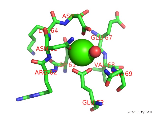
Calcium binding site 1 out of 2 in 4p2y
Go back to
Calcium binding site 1 out
of 2 in the Crystal Structure of the Human Rage Ectodomain (Fragment VC1C2) in Complex with Mouse S100A6
Mono view

Stereo pair view
Mono view
Stereo pair view
A full contact list of Calcium with other atoms in the Ca binding
site number 1 of Crystal Structure of the Human Rage Ectodomain (Fragment VC1C2) in Complex with Mouse S100A6 within 5.0Å range:
|
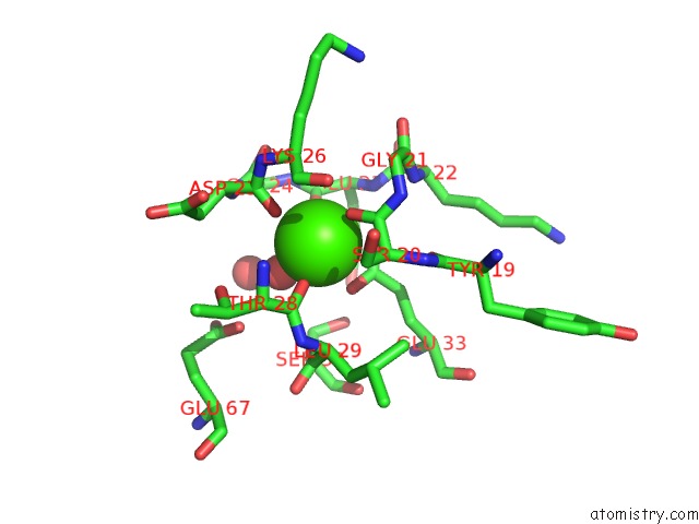
Calcium binding site 2 out of 2 in 4p2y
Go back to
Calcium binding site 2 out
of 2 in the Crystal Structure of the Human Rage Ectodomain (Fragment VC1C2) in Complex with Mouse S100A6
Mono view

Stereo pair view
Mono view
Stereo pair view
A full contact list of Calcium with other atoms in the Ca binding
site number 2 of Crystal Structure of the Human Rage Ectodomain (Fragment VC1C2) in Complex with Mouse S100A6 within 5.0Å range:
|
Reference:
L.Yatime,
C.Betzer,
S.Sirotkina,
P.H.Jensen,
G.R.Andersen.
The Structure of the Rage:S100A6 Complex Reveals A New Mode of Homodimerization For S100 Proteins To Be Published.
Page generated: Wed Jul 9 01:12:52 2025
Last articles
Mg in 6TRAMg in 6TR4
Mg in 6TR3
Mg in 6TMF
Mg in 6TQO
Mg in 6TQN
Mg in 6TQF
Mg in 6TQE
Mg in 6TQB
Mg in 6TQA