Calcium »
PDB 4p4i-4pjs »
4p5e »
Calcium in PDB 4p5e: Crystal Structure of Human DNPH1 (Rcl) with 6-Naphthyl-Purine- Riboside-Monophosphate
Protein crystallography data
The structure of Crystal Structure of Human DNPH1 (Rcl) with 6-Naphthyl-Purine- Riboside-Monophosphate, PDB code: 4p5e
was solved by
A.Padilla,
G.Labesse,
P.A.Kaminski,
with X-Ray Crystallography technique. A brief refinement statistics is given in the table below:
| Resolution Low / High (Å) | 47.36 / 1.35 |
| Space group | P 1 21 1 |
| Cell size a, b, c (Å), α, β, γ (°) | 48.974, 52.284, 54.454, 90.00, 104.77, 90.00 |
| R / Rfree (%) | 15.5 / 18 |
Calcium Binding Sites:
The binding sites of Calcium atom in the Crystal Structure of Human DNPH1 (Rcl) with 6-Naphthyl-Purine- Riboside-Monophosphate
(pdb code 4p5e). This binding sites where shown within
5.0 Angstroms radius around Calcium atom.
In total only one binding site of Calcium was determined in the Crystal Structure of Human DNPH1 (Rcl) with 6-Naphthyl-Purine- Riboside-Monophosphate, PDB code: 4p5e:
In total only one binding site of Calcium was determined in the Crystal Structure of Human DNPH1 (Rcl) with 6-Naphthyl-Purine- Riboside-Monophosphate, PDB code: 4p5e:
Calcium binding site 1 out of 1 in 4p5e
Go back to
Calcium binding site 1 out
of 1 in the Crystal Structure of Human DNPH1 (Rcl) with 6-Naphthyl-Purine- Riboside-Monophosphate

Mono view
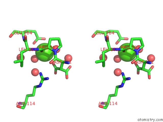
Stereo pair view
Mono view
Stereo pair view
A full contact list of Calcium with other atoms in the Ca binding
site number 1 of Crystal Structure of Human DNPH1 (Rcl) with 6-Naphthyl-Purine- Riboside-Monophosphate within 5.0Å range:
|
Reference:
C.Amiable,
J.Paoletti,
A.Haouz,
A.Padilla,
G.Labesse,
P.A.Kaminski,
S.Pochet.
6-(Hetero)Arylpurine Nucleotides As Inhibitors of the Oncogenic Target DNPH1: Synthesis, Structural Studies and Cytotoxic Activities. Eur.J.Med.Chem. V. 85C 418 2014.
ISSN: ISSN 0223-5234
PubMed: 25108359
DOI: 10.1016/J.EJMECH.2014.07.110
Page generated: Wed Jul 9 01:14:05 2025
ISSN: ISSN 0223-5234
PubMed: 25108359
DOI: 10.1016/J.EJMECH.2014.07.110
Last articles
Mg in 5MGYMg in 5MFM
Mg in 5MH1
Mg in 5MGA
Mg in 5M73
Mg in 5MFL
Mg in 5MF4
Mg in 5MF5
Mg in 5MCP
Mg in 5MDN