Calcium »
PDB 4p4i-4pjs »
4pc0 »
Calcium in PDB 4pc0: Structure of the Human RBAP48-MTA1(670-711) Complex
Protein crystallography data
The structure of Structure of the Human RBAP48-MTA1(670-711) Complex, PDB code: 4pc0
was solved by
S.S.M.Alqarni,
A.P.G.Silva,
J.P.Mackay,
E.D.Laue,
with X-Ray Crystallography technique. A brief refinement statistics is given in the table below:
| Resolution Low / High (Å) | 20.00 / 2.50 |
| Space group | P 1 21 1 |
| Cell size a, b, c (Å), α, β, γ (°) | 52.250, 123.230, 87.340, 90.00, 103.39, 90.00 |
| R / Rfree (%) | 20.4 / 25.9 |
Calcium Binding Sites:
The binding sites of Calcium atom in the Structure of the Human RBAP48-MTA1(670-711) Complex
(pdb code 4pc0). This binding sites where shown within
5.0 Angstroms radius around Calcium atom.
In total 2 binding sites of Calcium where determined in the Structure of the Human RBAP48-MTA1(670-711) Complex, PDB code: 4pc0:
Jump to Calcium binding site number: 1; 2;
In total 2 binding sites of Calcium where determined in the Structure of the Human RBAP48-MTA1(670-711) Complex, PDB code: 4pc0:
Jump to Calcium binding site number: 1; 2;
Calcium binding site 1 out of 2 in 4pc0
Go back to
Calcium binding site 1 out
of 2 in the Structure of the Human RBAP48-MTA1(670-711) Complex
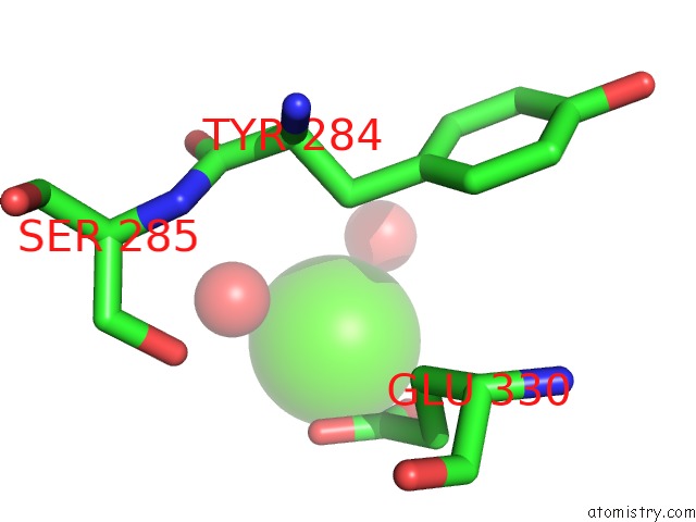
Mono view
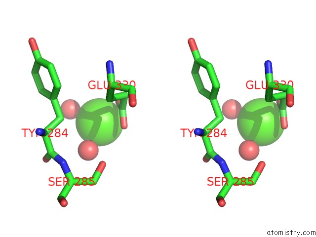
Stereo pair view
Mono view
Stereo pair view
A full contact list of Calcium with other atoms in the Ca binding
site number 1 of Structure of the Human RBAP48-MTA1(670-711) Complex within 5.0Å range:
|
Calcium binding site 2 out of 2 in 4pc0
Go back to
Calcium binding site 2 out
of 2 in the Structure of the Human RBAP48-MTA1(670-711) Complex
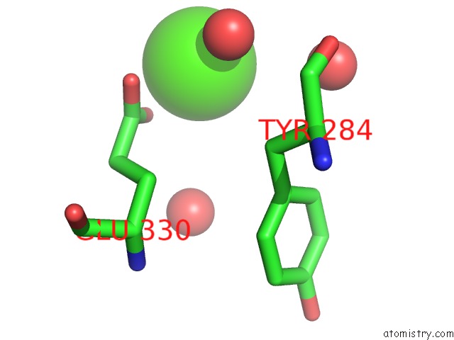
Mono view
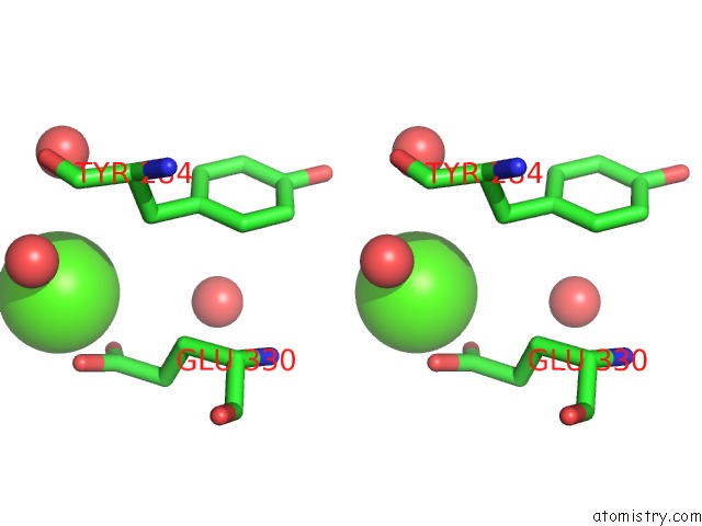
Stereo pair view
Mono view
Stereo pair view
A full contact list of Calcium with other atoms in the Ca binding
site number 2 of Structure of the Human RBAP48-MTA1(670-711) Complex within 5.0Å range:
|
Reference:
S.S.Alqarni,
A.Murthy,
W.Zhang,
M.R.Przewloka,
A.P.Silva,
A.A.Watson,
S.Lejon,
X.Y.Pei,
A.H.Smits,
S.L.Kloet,
H.Wang,
N.E.Shepherd,
P.H.Stokes,
G.A.Blobel,
M.Vermeulen,
D.M.Glover,
J.P.Mackay,
E.D.Laue.
Insight Into the Architecture of the Nurd Complex: Structure of the RBAP48-MTA1 Sub-Complex. J.Biol.Chem. V. 289 21844 2014.
ISSN: ISSN 0021-9258
PubMed: 24920672
DOI: 10.1074/JBC.M114.558940
Page generated: Wed Jul 9 01:16:13 2025
ISSN: ISSN 0021-9258
PubMed: 24920672
DOI: 10.1074/JBC.M114.558940
Last articles
Mg in 5MDLMg in 5MDK
Mg in 5MAC
Mg in 5MDJ
Mg in 5MBK
Mg in 5MAQ
Mg in 5MB9
Mg in 5M8G
Mg in 5M8D
Mg in 5M8B