Calcium »
PDB 4q1o-4qn4 »
4qb6 »
Calcium in PDB 4qb6: Structure of CBM35 in Complex with Aldouronic Acid
Enzymatic activity of Structure of CBM35 in Complex with Aldouronic Acid
All present enzymatic activity of Structure of CBM35 in Complex with Aldouronic Acid:
3.2.1.8;
3.2.1.8;
Protein crystallography data
The structure of Structure of CBM35 in Complex with Aldouronic Acid, PDB code: 4qb6
was solved by
M.A.Sainz-Polo,
J.Sanz-Aparicio,
with X-Ray Crystallography technique. A brief refinement statistics is given in the table below:
| Resolution Low / High (Å) | 51.51 / 1.35 |
| Space group | P 21 21 21 |
| Cell size a, b, c (Å), α, β, γ (°) | 39.780, 47.350, 103.030, 90.00, 90.00, 90.00 |
| R / Rfree (%) | 16.4 / 17.7 |
Calcium Binding Sites:
The binding sites of Calcium atom in the Structure of CBM35 in Complex with Aldouronic Acid
(pdb code 4qb6). This binding sites where shown within
5.0 Angstroms radius around Calcium atom.
In total 2 binding sites of Calcium where determined in the Structure of CBM35 in Complex with Aldouronic Acid, PDB code: 4qb6:
Jump to Calcium binding site number: 1; 2;
In total 2 binding sites of Calcium where determined in the Structure of CBM35 in Complex with Aldouronic Acid, PDB code: 4qb6:
Jump to Calcium binding site number: 1; 2;
Calcium binding site 1 out of 2 in 4qb6
Go back to
Calcium binding site 1 out
of 2 in the Structure of CBM35 in Complex with Aldouronic Acid
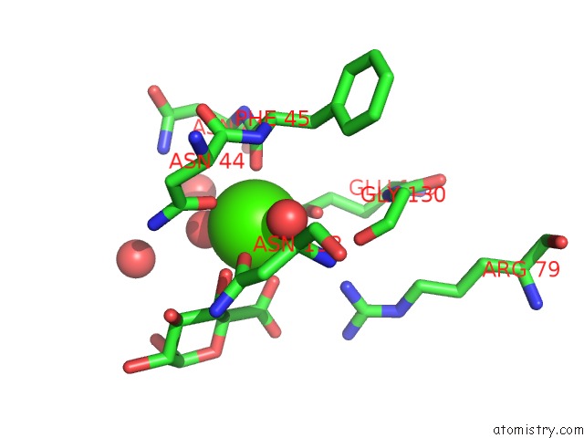
Mono view
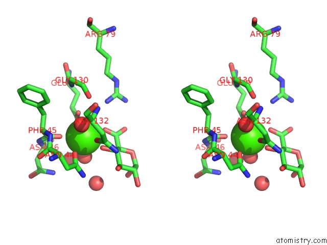
Stereo pair view
Mono view
Stereo pair view
A full contact list of Calcium with other atoms in the Ca binding
site number 1 of Structure of CBM35 in Complex with Aldouronic Acid within 5.0Å range:
|
Calcium binding site 2 out of 2 in 4qb6
Go back to
Calcium binding site 2 out
of 2 in the Structure of CBM35 in Complex with Aldouronic Acid
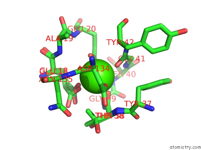
Mono view
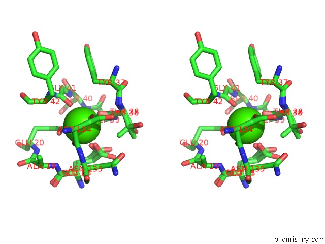
Stereo pair view
Mono view
Stereo pair view
A full contact list of Calcium with other atoms in the Ca binding
site number 2 of Structure of CBM35 in Complex with Aldouronic Acid within 5.0Å range:
|
Reference:
M.A.Sainz-Polo,
S.V.Valenzuela,
B.Gonzalez,
F.I.Pastor,
J.Sanz-Aparicio.
Structural Analysis of Glucuronoxylan Specific XYN30D and Its Attached CBM35 Domain Give Insights Into the Role of Modularity in Specificity. J.Biol.Chem. 2014.
ISSN: ESSN 1083-351X
PubMed: 25202007
DOI: 10.1074/JBC.M114.597732
Page generated: Wed Jul 9 01:42:38 2025
ISSN: ESSN 1083-351X
PubMed: 25202007
DOI: 10.1074/JBC.M114.597732
Last articles
Fe in 2YXOFe in 2YRS
Fe in 2YXC
Fe in 2YNM
Fe in 2YVJ
Fe in 2YP1
Fe in 2YU2
Fe in 2YU1
Fe in 2YQB
Fe in 2YOO