Calcium »
PDB 4qn5-4r90 »
4qo5 »
Calcium in PDB 4qo5: Hypothetical Multiheme Protein
Protein crystallography data
The structure of Hypothetical Multiheme Protein, PDB code: 4qo5
was solved by
C.Rajendran,
with X-Ray Crystallography technique. A brief refinement statistics is given in the table below:
| Resolution Low / High (Å) | 43.73 / 1.70 |
| Space group | H 3 2 |
| Cell size a, b, c (Å), α, β, γ (°) | 136.440, 136.440, 214.900, 90.00, 90.00, 120.00 |
| R / Rfree (%) | 16.5 / 20.1 |
Other elements in 4qo5:
The structure of Hypothetical Multiheme Protein also contains other interesting chemical elements:
| Iron | (Fe) | 8 atoms |
Calcium Binding Sites:
The binding sites of Calcium atom in the Hypothetical Multiheme Protein
(pdb code 4qo5). This binding sites where shown within
5.0 Angstroms radius around Calcium atom.
In total only one binding site of Calcium was determined in the Hypothetical Multiheme Protein, PDB code: 4qo5:
In total only one binding site of Calcium was determined in the Hypothetical Multiheme Protein, PDB code: 4qo5:
Calcium binding site 1 out of 1 in 4qo5
Go back to
Calcium binding site 1 out
of 1 in the Hypothetical Multiheme Protein
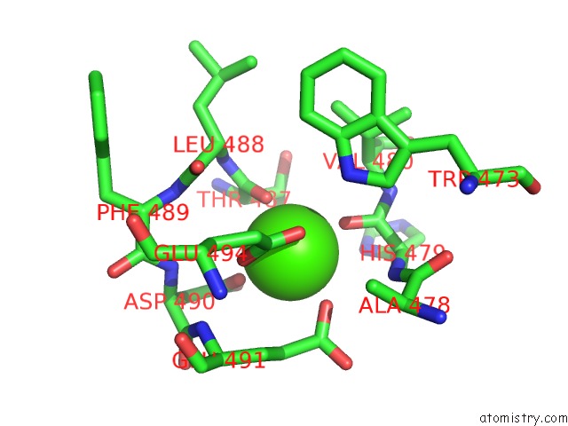
Mono view
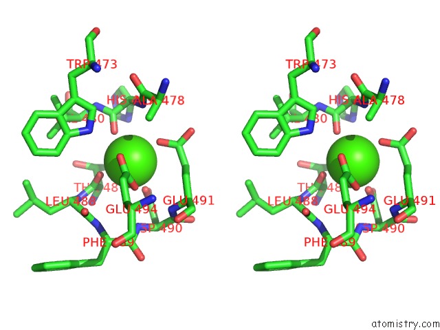
Stereo pair view
Mono view
Stereo pair view
A full contact list of Calcium with other atoms in the Ca binding
site number 1 of Hypothetical Multiheme Protein within 5.0Å range:
|
Reference:
K.Parey,
A.J.Fielding,
M.Sorgel,
R.Rachel,
H.Huber,
C.Ziegler,
C.Rajendran.
In Meso Crystal Structure of A Novel Membrane-Associated Octaheme Cytochrome C From the Crenarchaeon Ignicoccus Hospitalis. Febs J. V. 283 3807 2016.
ISSN: ISSN 1742-464X
PubMed: 27586496
DOI: 10.1111/FEBS.13870
Page generated: Wed Jul 9 01:46:37 2025
ISSN: ISSN 1742-464X
PubMed: 27586496
DOI: 10.1111/FEBS.13870
Last articles
Mg in 5NOLMg in 5NOJ
Mg in 5NOG
Mg in 5NO9
Mg in 5NNW
Mg in 5NO1
Mg in 5NLM
Mg in 5NMX
Mg in 5NMW
Mg in 5NH7