Calcium »
PDB 4qn5-4r90 »
4qpj »
Calcium in PDB 4qpj: 2.7 Angstrom Structure of A Phosphotransferase in Complex with A Receiver Domain
Protein crystallography data
The structure of 2.7 Angstrom Structure of A Phosphotransferase in Complex with A Receiver Domain, PDB code: 4qpj
was solved by
J.W.Willett,
J.Herrou,
S.Crosson,
with X-Ray Crystallography technique. A brief refinement statistics is given in the table below:
| Resolution Low / High (Å) | 17.88 / 2.74 |
| Space group | P 32 2 1 |
| Cell size a, b, c (Å), α, β, γ (°) | 124.951, 124.951, 136.332, 90.00, 90.00, 120.00 |
| R / Rfree (%) | 17.5 / 22.2 |
Other elements in 4qpj:
The structure of 2.7 Angstrom Structure of A Phosphotransferase in Complex with A Receiver Domain also contains other interesting chemical elements:
| Magnesium | (Mg) | 1 atom |
| Chlorine | (Cl) | 1 atom |
| Sodium | (Na) | 1 atom |
Calcium Binding Sites:
The binding sites of Calcium atom in the 2.7 Angstrom Structure of A Phosphotransferase in Complex with A Receiver Domain
(pdb code 4qpj). This binding sites where shown within
5.0 Angstroms radius around Calcium atom.
In total 3 binding sites of Calcium where determined in the 2.7 Angstrom Structure of A Phosphotransferase in Complex with A Receiver Domain, PDB code: 4qpj:
Jump to Calcium binding site number: 1; 2; 3;
In total 3 binding sites of Calcium where determined in the 2.7 Angstrom Structure of A Phosphotransferase in Complex with A Receiver Domain, PDB code: 4qpj:
Jump to Calcium binding site number: 1; 2; 3;
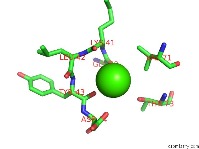
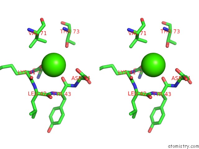
Calcium binding site 1 out of 3 in 4qpj
Go back to
Calcium binding site 1 out
of 3 in the 2.7 Angstrom Structure of A Phosphotransferase in Complex with A Receiver Domain
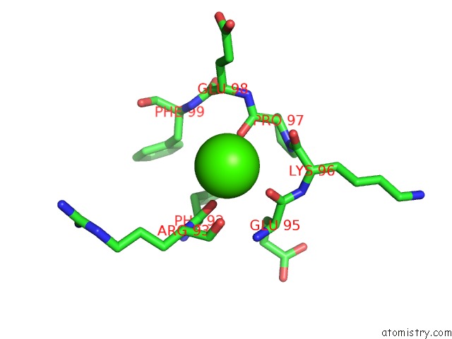
Mono view
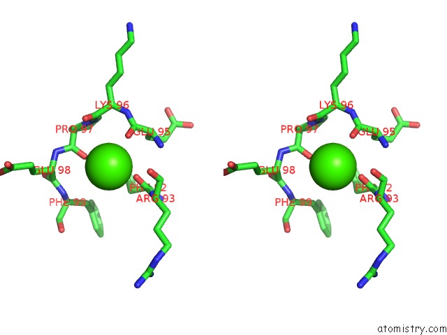
Stereo pair view
Mono view
Stereo pair view
A full contact list of Calcium with other atoms in the Ca binding
site number 1 of 2.7 Angstrom Structure of A Phosphotransferase in Complex with A Receiver Domain within 5.0Å range:
|
Calcium binding site 2 out of 3 in 4qpj
Go back to
Calcium binding site 2 out
of 3 in the 2.7 Angstrom Structure of A Phosphotransferase in Complex with A Receiver Domain
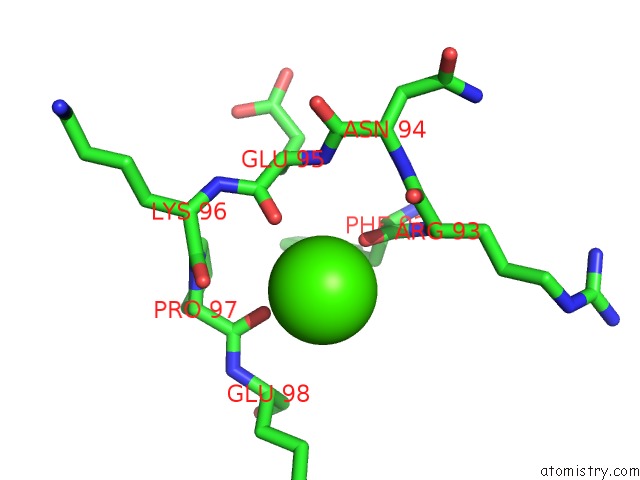
Mono view
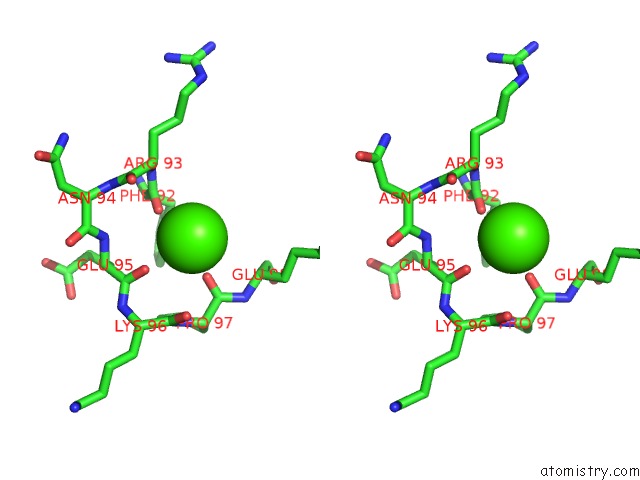
Stereo pair view
Mono view
Stereo pair view
A full contact list of Calcium with other atoms in the Ca binding
site number 2 of 2.7 Angstrom Structure of A Phosphotransferase in Complex with A Receiver Domain within 5.0Å range:
|
Calcium binding site 3 out of 3 in 4qpj
Go back to
Calcium binding site 3 out
of 3 in the 2.7 Angstrom Structure of A Phosphotransferase in Complex with A Receiver Domain
Mono view
Stereo pair view
Mono view
Stereo pair view
A full contact list of Calcium with other atoms in the Ca binding
site number 3 of 2.7 Angstrom Structure of A Phosphotransferase in Complex with A Receiver Domain within 5.0Å range:
|
Reference:
J.W.Willett,
J.Herrou,
A.Briegel,
G.Rotskoff,
S.Crosson.
Structural Asymmetry in A Conserved Signaling System That Regulates Division, Replication, and Virulence of An Intracellular Pathogen. Proc.Natl.Acad.Sci.Usa V. 112 E3709 2015.
ISSN: ISSN 0027-8424
PubMed: 26124143
DOI: 10.1073/PNAS.1503118112
Page generated: Wed Jul 9 01:46:44 2025
ISSN: ISSN 0027-8424
PubMed: 26124143
DOI: 10.1073/PNAS.1503118112
Last articles
Fe in 6ONRFe in 6ONQ
Fe in 6ONK
Fe in 6ONG
Fe in 6ON3
Fe in 6ON1
Fe in 6OJW
Fe in 6OM5
Fe in 6OFD
Fe in 6OF5