Calcium »
PDB 4qn5-4r90 »
4r5k »
Calcium in PDB 4r5k: Crystal Structure of the Dnak C-Terminus (Dnak-Sbd-B)
Protein crystallography data
The structure of Crystal Structure of the Dnak C-Terminus (Dnak-Sbd-B), PDB code: 4r5k
was solved by
J.I.Leu,
P.Zhang,
M.E.Murphy,
R.Marmorstein,
D.L.George,
with X-Ray Crystallography technique. A brief refinement statistics is given in the table below:
| Resolution Low / High (Å) | 41.24 / 1.75 |
| Space group | P 1 |
| Cell size a, b, c (Å), α, β, γ (°) | 43.636, 44.827, 63.174, 77.30, 78.14, 72.96 |
| R / Rfree (%) | 17.6 / 21.8 |
Calcium Binding Sites:
The binding sites of Calcium atom in the Crystal Structure of the Dnak C-Terminus (Dnak-Sbd-B)
(pdb code 4r5k). This binding sites where shown within
5.0 Angstroms radius around Calcium atom.
In total only one binding site of Calcium was determined in the Crystal Structure of the Dnak C-Terminus (Dnak-Sbd-B), PDB code: 4r5k:
In total only one binding site of Calcium was determined in the Crystal Structure of the Dnak C-Terminus (Dnak-Sbd-B), PDB code: 4r5k:
Calcium binding site 1 out of 1 in 4r5k
Go back to
Calcium binding site 1 out
of 1 in the Crystal Structure of the Dnak C-Terminus (Dnak-Sbd-B)
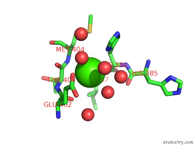
Mono view
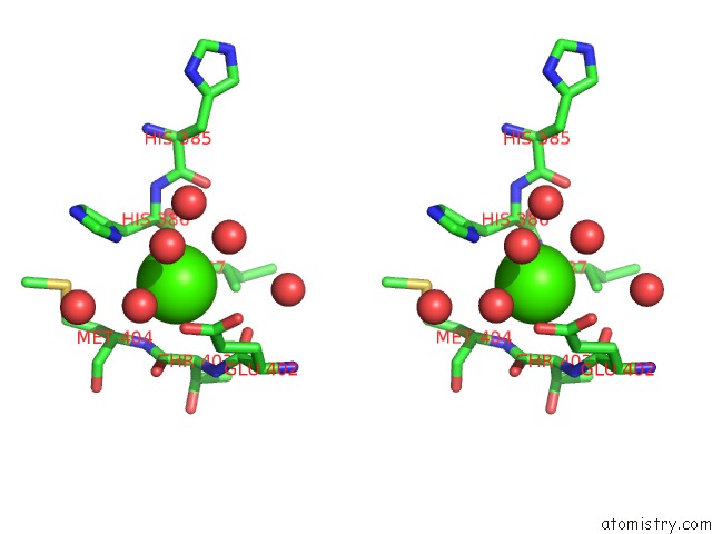
Stereo pair view
Mono view
Stereo pair view
A full contact list of Calcium with other atoms in the Ca binding
site number 1 of Crystal Structure of the Dnak C-Terminus (Dnak-Sbd-B) within 5.0Å range:
|
Reference:
J.I.Leu,
P.Zhang,
M.E.Murphy,
R.Marmorstein,
D.L.George.
Structural Basis For the Inhibition of HSP70 and Dnak Chaperones By Small-Molecule Targeting of A C-Terminal Allosteric Pocket. Acs Chem.Biol. 2014.
ISSN: ESSN 1554-8937
PubMed: 25148104
DOI: 10.1021/CB500236Y
Page generated: Wed Jul 9 01:51:00 2025
ISSN: ESSN 1554-8937
PubMed: 25148104
DOI: 10.1021/CB500236Y
Last articles
Mg in 3LEEMg in 3LG5
Mg in 3LER
Mg in 3LA5
Mg in 3LDV
Mg in 3LCB
Mg in 3LDL
Mg in 3LC8
Mg in 3LBN
Mg in 3LC6