Calcium »
PDB 4tx8-4uix »
4uap »
Calcium in PDB 4uap: X-Ray Structure of GH31 CBM32-2 Bound to Galnac
Protein crystallography data
The structure of X-Ray Structure of GH31 CBM32-2 Bound to Galnac, PDB code: 4uap
was solved by
J.M.Grondin,
K.Abe,
A.B.Boraston,
S.P.Smith,
with X-Ray Crystallography technique. A brief refinement statistics is given in the table below:
| Resolution Low / High (Å) | 38.41 / 2.00 |
| Space group | P 2 21 21 |
| Cell size a, b, c (Å), α, β, γ (°) | 48.300, 84.750, 86.170, 90.00, 90.00, 90.00 |
| R / Rfree (%) | 14.5 / 19.2 |
Calcium Binding Sites:
The binding sites of Calcium atom in the X-Ray Structure of GH31 CBM32-2 Bound to Galnac
(pdb code 4uap). This binding sites where shown within
5.0 Angstroms radius around Calcium atom.
In total 2 binding sites of Calcium where determined in the X-Ray Structure of GH31 CBM32-2 Bound to Galnac, PDB code: 4uap:
Jump to Calcium binding site number: 1; 2;
In total 2 binding sites of Calcium where determined in the X-Ray Structure of GH31 CBM32-2 Bound to Galnac, PDB code: 4uap:
Jump to Calcium binding site number: 1; 2;
Calcium binding site 1 out of 2 in 4uap
Go back to
Calcium binding site 1 out
of 2 in the X-Ray Structure of GH31 CBM32-2 Bound to Galnac
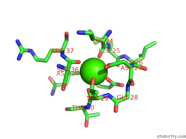
Mono view
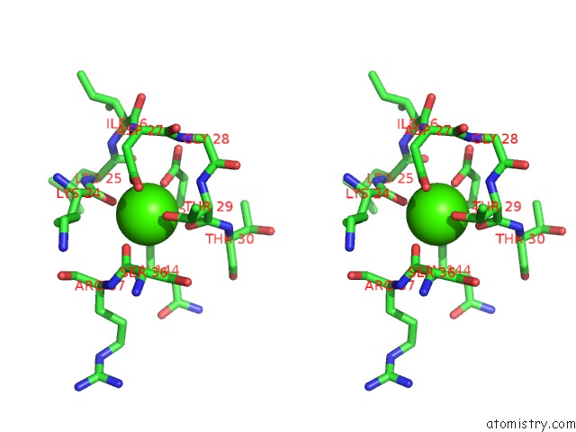
Stereo pair view
Mono view
Stereo pair view
A full contact list of Calcium with other atoms in the Ca binding
site number 1 of X-Ray Structure of GH31 CBM32-2 Bound to Galnac within 5.0Å range:
|
Calcium binding site 2 out of 2 in 4uap
Go back to
Calcium binding site 2 out
of 2 in the X-Ray Structure of GH31 CBM32-2 Bound to Galnac
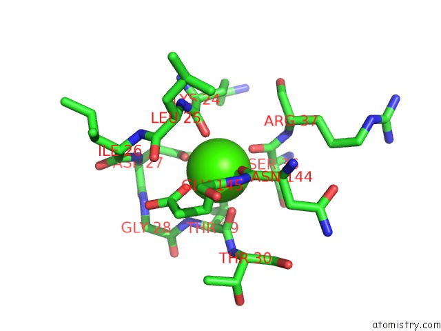
Mono view
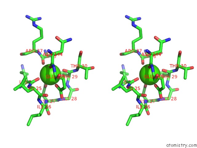
Stereo pair view
Mono view
Stereo pair view
A full contact list of Calcium with other atoms in the Ca binding
site number 2 of X-Ray Structure of GH31 CBM32-2 Bound to Galnac within 5.0Å range:
|
Reference:
J.M.Grondin,
D.Duan,
A.C.Kirlin,
K.T.Abe,
S.Chitayat,
H.L.Spencer,
C.Spencer,
A.Campigotto,
S.Houliston,
C.H.Arrowsmith,
J.S.Allingham,
A.B.Boraston,
S.P.Smith.
Diverse Modes of Galacto-Specific Carbohydrate Recognition By A Family 31 Glycoside Hydrolase From Clostridium Perfringens. Plos One V. 12 71606 2017.
ISSN: ESSN 1932-6203
PubMed: 28158290
DOI: 10.1371/JOURNAL.PONE.0171606
Page generated: Wed Jul 9 02:15:26 2025
ISSN: ESSN 1932-6203
PubMed: 28158290
DOI: 10.1371/JOURNAL.PONE.0171606
Last articles
K in 3FWPK in 3G4S
K in 3FWF
K in 3FWG
K in 3FWJ
K in 3FWI
K in 3FTM
K in 3FTF
K in 3FPB
K in 3FN2