Calcium »
PDB 4uj7-4w9y »
4ur9 »
Calcium in PDB 4ur9: Structure of Ligand Bound Glycosylhydrolase
Enzymatic activity of Structure of Ligand Bound Glycosylhydrolase
All present enzymatic activity of Structure of Ligand Bound Glycosylhydrolase:
3.2.1.169;
3.2.1.169;
Protein crystallography data
The structure of Structure of Ligand Bound Glycosylhydrolase, PDB code: 4ur9
was solved by
J.F.Darby,
J.Landstroem,
C.Roth,
Y.He,
M.Schultz,
G.J.Davies,
R.E.Hubbard,
with X-Ray Crystallography technique. A brief refinement statistics is given in the table below:
| Resolution Low / High (Å) | 48.786 / 2.20 |
| Space group | P 2 21 21 |
| Cell size a, b, c (Å), α, β, γ (°) | 51.486, 162.735, 223.179, 90.00, 90.00, 90.00 |
| R / Rfree (%) | 22.15 / 25.2 |
Calcium Binding Sites:
The binding sites of Calcium atom in the Structure of Ligand Bound Glycosylhydrolase
(pdb code 4ur9). This binding sites where shown within
5.0 Angstroms radius around Calcium atom.
In total only one binding site of Calcium was determined in the Structure of Ligand Bound Glycosylhydrolase, PDB code: 4ur9:
In total only one binding site of Calcium was determined in the Structure of Ligand Bound Glycosylhydrolase, PDB code: 4ur9:
Calcium binding site 1 out of 1 in 4ur9
Go back to
Calcium binding site 1 out
of 1 in the Structure of Ligand Bound Glycosylhydrolase
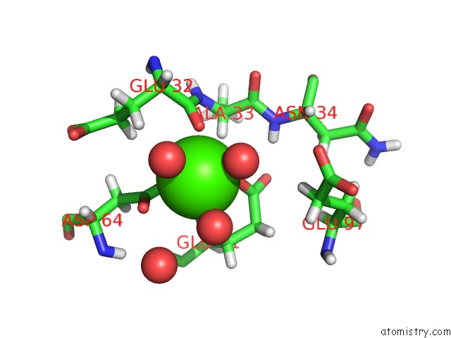
Mono view
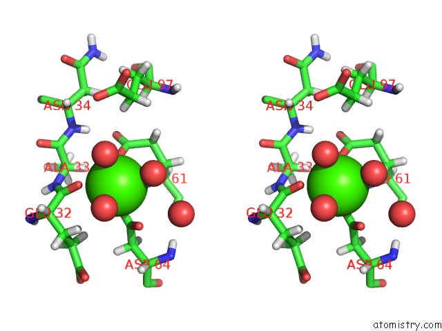
Stereo pair view
Mono view
Stereo pair view
A full contact list of Calcium with other atoms in the Ca binding
site number 1 of Structure of Ligand Bound Glycosylhydrolase within 5.0Å range:
|
Reference:
J.F.Darby,
J.Landstrom,
C.Roth,
Y.He,
G.J.Davies,
R.E.Hubbard.
Discovery of Selective Small-Molecule Activators of A Bacterial Glycoside Hydrolase. Angew.Chem.Int.Ed.Engl. V. 53 13419 2014.
ISSN: ISSN 1433-7851
PubMed: 25291993
DOI: 10.1002/ANIE.201407081
Page generated: Wed Jul 9 02:23:45 2025
ISSN: ISSN 1433-7851
PubMed: 25291993
DOI: 10.1002/ANIE.201407081
Last articles
Mg in 4GNRMg in 4GOJ
Mg in 4GOK
Mg in 4GKK
Mg in 4GKJ
Mg in 4GMJ
Mg in 4GNK
Mg in 4GNI
Mg in 4GN0
Mg in 4GMX