Calcium »
PDB 4uj7-4w9y »
4usv »
Calcium in PDB 4usv: Crystal Structure of Human Soluble Adenylyl Cyclase with Pyrophosphate Resulting From Soaking with Atp and Calcium
Enzymatic activity of Crystal Structure of Human Soluble Adenylyl Cyclase with Pyrophosphate Resulting From Soaking with Atp and Calcium
All present enzymatic activity of Crystal Structure of Human Soluble Adenylyl Cyclase with Pyrophosphate Resulting From Soaking with Atp and Calcium:
4.6.1.1;
4.6.1.1;
Protein crystallography data
The structure of Crystal Structure of Human Soluble Adenylyl Cyclase with Pyrophosphate Resulting From Soaking with Atp and Calcium, PDB code: 4usv
was solved by
S.Kleinboelting,
C.Steegborn,
with X-Ray Crystallography technique. A brief refinement statistics is given in the table below:
| Resolution Low / High (Å) | 87.36 / 2.00 |
| Space group | P 63 |
| Cell size a, b, c (Å), α, β, γ (°) | 100.870, 100.870, 97.150, 90.00, 90.00, 120.00 |
| R / Rfree (%) | 15.994 / 20.893 |
Other elements in 4usv:
The structure of Crystal Structure of Human Soluble Adenylyl Cyclase with Pyrophosphate Resulting From Soaking with Atp and Calcium also contains other interesting chemical elements:
| Chlorine | (Cl) | 1 atom |
Calcium Binding Sites:
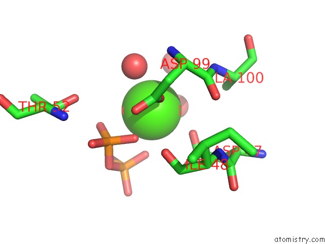
The binding sites of Calcium atom in the Crystal Structure of Human Soluble Adenylyl Cyclase with Pyrophosphate Resulting From Soaking with Atp and Calcium
(pdb code 4usv). This binding sites where shown within
5.0 Angstroms radius around Calcium atom.
In total only one binding site of Calcium was determined in the Crystal Structure of Human Soluble Adenylyl Cyclase with Pyrophosphate Resulting From Soaking with Atp and Calcium, PDB code: 4usv:
In total only one binding site of Calcium was determined in the Crystal Structure of Human Soluble Adenylyl Cyclase with Pyrophosphate Resulting From Soaking with Atp and Calcium, PDB code: 4usv:
Calcium binding site 1 out of 1 in 4usv
Go back to
Calcium binding site 1 out
of 1 in the Crystal Structure of Human Soluble Adenylyl Cyclase with Pyrophosphate Resulting From Soaking with Atp and Calcium
Mono view

Stereo pair view
Mono view
Stereo pair view
A full contact list of Calcium with other atoms in the Ca binding
site number 1 of Crystal Structure of Human Soluble Adenylyl Cyclase with Pyrophosphate Resulting From Soaking with Atp and Calcium within 5.0Å range:
|
Reference:
S.Kleinbolting,
J.Van Den Heuvel,
C.Steegborn.
Structural Analysis of Human Soluble Adenylyl Cyclase and Crystal Structures of Its Nucleotide Complexes - Implications For Cyclase Catalysis and Evolution. Febs J. V. 281 4151 2014.
ISSN: ISSN 1742-464X
PubMed: 25040695
DOI: 10.1111/FEBS.12913
Page generated: Wed Jul 9 02:24:53 2025
ISSN: ISSN 1742-464X
PubMed: 25040695
DOI: 10.1111/FEBS.12913
Last articles
Mg in 1YL7Mg in 1YL6
Mg in 1YL5
Mg in 1YKV
Mg in 1YKQ
Mg in 1YIT
Mg in 1YIJ
Mg in 1YJF
Mg in 1YI2
Mg in 1YJ2