Calcium »
PDB 4wa3-4wn0 »
4wi0 »
Calcium in PDB 4wi0: Crystal Structure of COH3SCAB-XDOC_M2SCAA Complex: A C-Terminal Interface Mutant of Type II Cohesin-X-Dockerin Complex From Acetivibrio Cellulolyticus
Protein crystallography data
The structure of Crystal Structure of COH3SCAB-XDOC_M2SCAA Complex: A C-Terminal Interface Mutant of Type II Cohesin-X-Dockerin Complex From Acetivibrio Cellulolyticus, PDB code: 4wi0
was solved by
V.D.Alves,
K.Cameron,
S.H.Najmudin,
C.M.G.A.Fontes,
with X-Ray Crystallography technique. A brief refinement statistics is given in the table below:
| Resolution Low / High (Å) | 63.71 / 1.93 |
| Space group | P 21 21 21 |
| Cell size a, b, c (Å), α, β, γ (°) | 38.540, 88.300, 92.020, 90.00, 90.00, 90.00 |
| R / Rfree (%) | 19.7 / 24.1 |
Calcium Binding Sites:
The binding sites of Calcium atom in the Crystal Structure of COH3SCAB-XDOC_M2SCAA Complex: A C-Terminal Interface Mutant of Type II Cohesin-X-Dockerin Complex From Acetivibrio Cellulolyticus
(pdb code 4wi0). This binding sites where shown within
5.0 Angstroms radius around Calcium atom.
In total 2 binding sites of Calcium where determined in the Crystal Structure of COH3SCAB-XDOC_M2SCAA Complex: A C-Terminal Interface Mutant of Type II Cohesin-X-Dockerin Complex From Acetivibrio Cellulolyticus, PDB code: 4wi0:
Jump to Calcium binding site number: 1; 2;
In total 2 binding sites of Calcium where determined in the Crystal Structure of COH3SCAB-XDOC_M2SCAA Complex: A C-Terminal Interface Mutant of Type II Cohesin-X-Dockerin Complex From Acetivibrio Cellulolyticus, PDB code: 4wi0:
Jump to Calcium binding site number: 1; 2;
Calcium binding site 1 out of 2 in 4wi0
Go back to
Calcium binding site 1 out
of 2 in the Crystal Structure of COH3SCAB-XDOC_M2SCAA Complex: A C-Terminal Interface Mutant of Type II Cohesin-X-Dockerin Complex From Acetivibrio Cellulolyticus
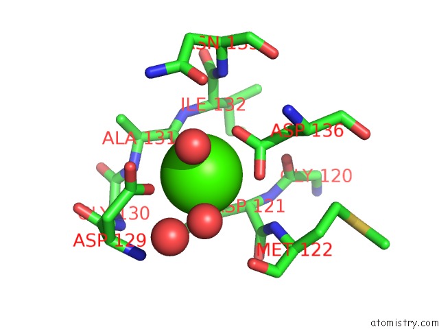
Mono view
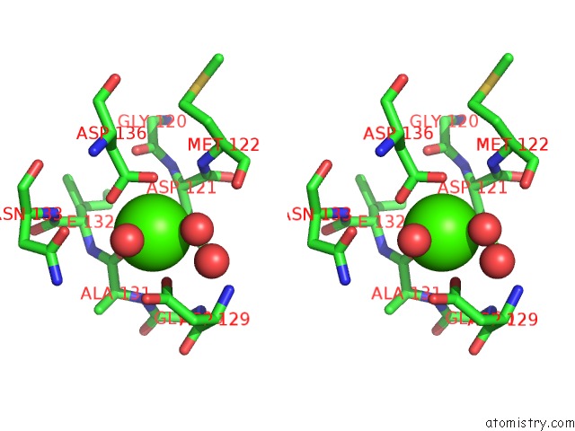
Stereo pair view
Mono view
Stereo pair view
A full contact list of Calcium with other atoms in the Ca binding
site number 1 of Crystal Structure of COH3SCAB-XDOC_M2SCAA Complex: A C-Terminal Interface Mutant of Type II Cohesin-X-Dockerin Complex From Acetivibrio Cellulolyticus within 5.0Å range:
|
Calcium binding site 2 out of 2 in 4wi0
Go back to
Calcium binding site 2 out
of 2 in the Crystal Structure of COH3SCAB-XDOC_M2SCAA Complex: A C-Terminal Interface Mutant of Type II Cohesin-X-Dockerin Complex From Acetivibrio Cellulolyticus
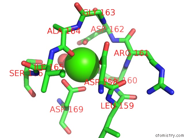
Mono view
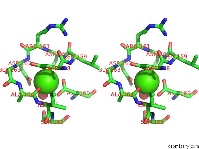
Stereo pair view
Mono view
Stereo pair view
A full contact list of Calcium with other atoms in the Ca binding
site number 2 of Crystal Structure of COH3SCAB-XDOC_M2SCAA Complex: A C-Terminal Interface Mutant of Type II Cohesin-X-Dockerin Complex From Acetivibrio Cellulolyticus within 5.0Å range:
|
Reference:
V.D.Alves,
K.Cameron,
S.H.Najmudin,
C.M.G.A.Fontes.
Crystal Structure of COH3SCAB-XDOC_M1SCAA Complex: A C-Terminal Interface Mutant of Type II Cohesin-X-Dockerin Complex From Acetivibrio Cellulolyticus To Be Published.
Page generated: Wed Jul 9 02:34:13 2025
Last articles
Mg in 2UU7Mg in 2UAG
Mg in 2UKD
Mg in 2SHK
Mg in 2TPS
Mg in 2TRT
Mg in 2TRA
Mg in 2RMK
Mg in 2RUS
Mg in 2TCT