Calcium »
PDB 4wa3-4wn0 »
4wit »
Calcium in PDB 4wit: TMEM16 Lipid Scramblase in Crystal Form 2
Protein crystallography data
The structure of TMEM16 Lipid Scramblase in Crystal Form 2, PDB code: 4wit
was solved by
R.Dutzler,
J.D.Brunner,
N.K.Lim,
S.Schenck,
with X-Ray Crystallography technique. A brief refinement statistics is given in the table below:
| Resolution Low / High (Å) | 15.00 / 3.40 |
| Space group | P 21 21 21 |
| Cell size a, b, c (Å), α, β, γ (°) | 115.940, 127.240, 180.110, 90.00, 90.00, 90.00 |
| R / Rfree (%) | 24.8 / 29.2 |
Calcium Binding Sites:
The binding sites of Calcium atom in the TMEM16 Lipid Scramblase in Crystal Form 2
(pdb code 4wit). This binding sites where shown within
5.0 Angstroms radius around Calcium atom.
In total 4 binding sites of Calcium where determined in the TMEM16 Lipid Scramblase in Crystal Form 2, PDB code: 4wit:
Jump to Calcium binding site number: 1; 2; 3; 4;
In total 4 binding sites of Calcium where determined in the TMEM16 Lipid Scramblase in Crystal Form 2, PDB code: 4wit:
Jump to Calcium binding site number: 1; 2; 3; 4;
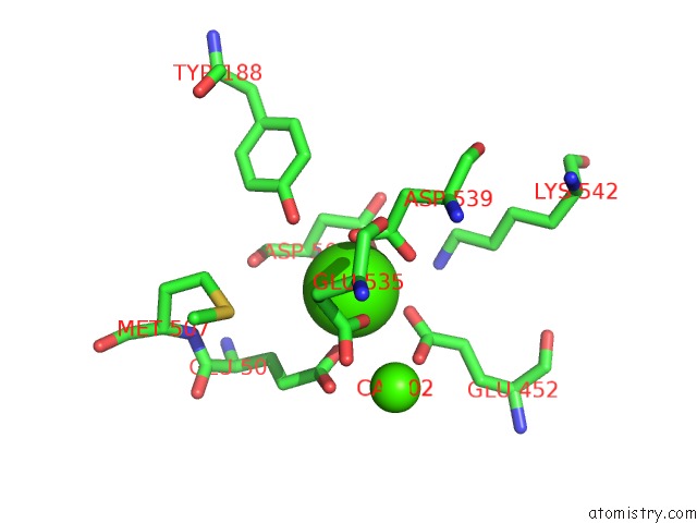
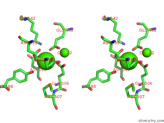
Calcium binding site 1 out of 4 in 4wit
Go back to
Calcium binding site 1 out
of 4 in the TMEM16 Lipid Scramblase in Crystal Form 2
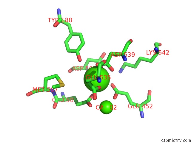
Mono view
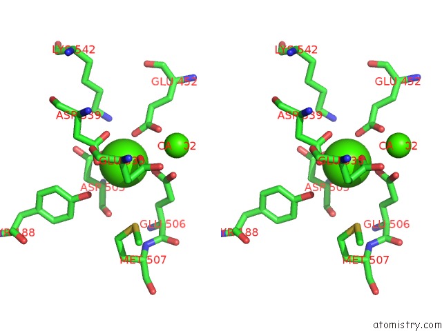
Stereo pair view
Mono view
Stereo pair view
A full contact list of Calcium with other atoms in the Ca binding
site number 1 of TMEM16 Lipid Scramblase in Crystal Form 2 within 5.0Å range:
|
Calcium binding site 2 out of 4 in 4wit
Go back to
Calcium binding site 2 out
of 4 in the TMEM16 Lipid Scramblase in Crystal Form 2
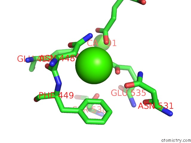
Mono view
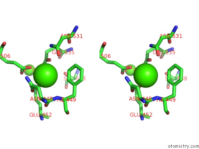
Stereo pair view
Mono view
Stereo pair view
A full contact list of Calcium with other atoms in the Ca binding
site number 2 of TMEM16 Lipid Scramblase in Crystal Form 2 within 5.0Å range:
|
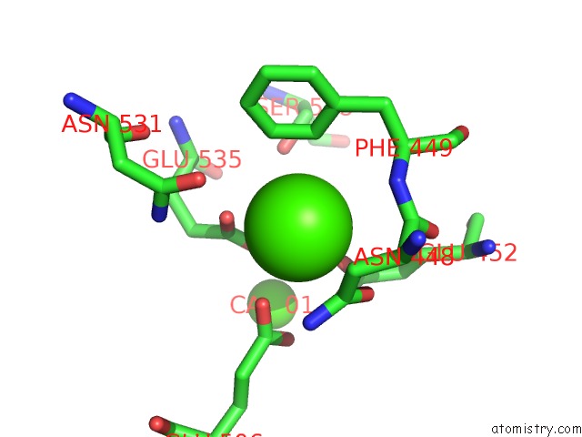
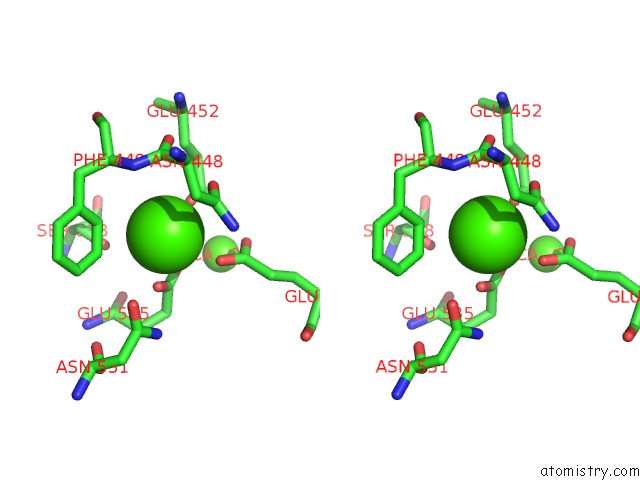
Calcium binding site 3 out of 4 in 4wit
Go back to
Calcium binding site 3 out
of 4 in the TMEM16 Lipid Scramblase in Crystal Form 2
Mono view
Stereo pair view
Mono view
Stereo pair view
A full contact list of Calcium with other atoms in the Ca binding
site number 3 of TMEM16 Lipid Scramblase in Crystal Form 2 within 5.0Å range:
|
Calcium binding site 4 out of 4 in 4wit
Go back to
Calcium binding site 4 out
of 4 in the TMEM16 Lipid Scramblase in Crystal Form 2
Mono view
Stereo pair view
Mono view
Stereo pair view
A full contact list of Calcium with other atoms in the Ca binding
site number 4 of TMEM16 Lipid Scramblase in Crystal Form 2 within 5.0Å range:
|
Reference:
J.D.Brunner,
N.K.Lim,
S.Schenck,
A.Duerst,
R.Dutzler.
X-Ray Structure of A Calcium-Activated TMEM16 Lipid Scramblase. Nature 2014.
ISSN: ESSN 1476-4687
PubMed: 25383531
DOI: 10.1038/NATURE13984
Page generated: Wed Jul 9 02:34:48 2025
ISSN: ESSN 1476-4687
PubMed: 25383531
DOI: 10.1038/NATURE13984
Last articles
K in 7QR0K in 7QR1
K in 7QQZ
K in 7QQX
K in 7QQW
K in 7QQU
K in 7QQV
K in 7QQT
K in 7QQR
K in 7QQS