Atomistry » Calcium » PDB 4wnb-4x8d » 4wyb
Atomistry »
  Calcium »
    PDB 4wnb-4x8d »
      4wyb »

Calcium in PDB 4wyb: Structure of the BUD6 Flank Domain in Complex with Actin

Protein crystallography data

The structure of Structure of the BUD6 Flank Domain in Complex with Actin, PDB code: 4wyb was solved by M.J.Eck, E.Park, W.Zheng, with X-Ray Crystallography technique. A brief refinement statistics is given in the table below:

Resolution Low / High (Å) 45.42 / 3.49
Space group P 32
Cell size a, b, c (Å), α, β, γ (°) 138.753, 138.753, 356.651, 90.00, 90.00, 120.00
R / Rfree (%) 21.2 / 25.8

Calcium Binding Sites:

Pages:

>>> Page 1 <<< Page 2, Binding sites: 11 - 12;

Binding sites:

The binding sites of Calcium atom in the Structure of the BUD6 Flank Domain in Complex with Actin (pdb code 4wyb). This binding sites where shown within 5.0 Angstroms radius around Calcium atom.
In total 12 binding sites of Calcium where determined in the Structure of the BUD6 Flank Domain in Complex with Actin, PDB code: 4wyb:
Jump to Calcium binding site number: 1; 2; 3; 4; 5; 6; 7; 8; 9; 10;

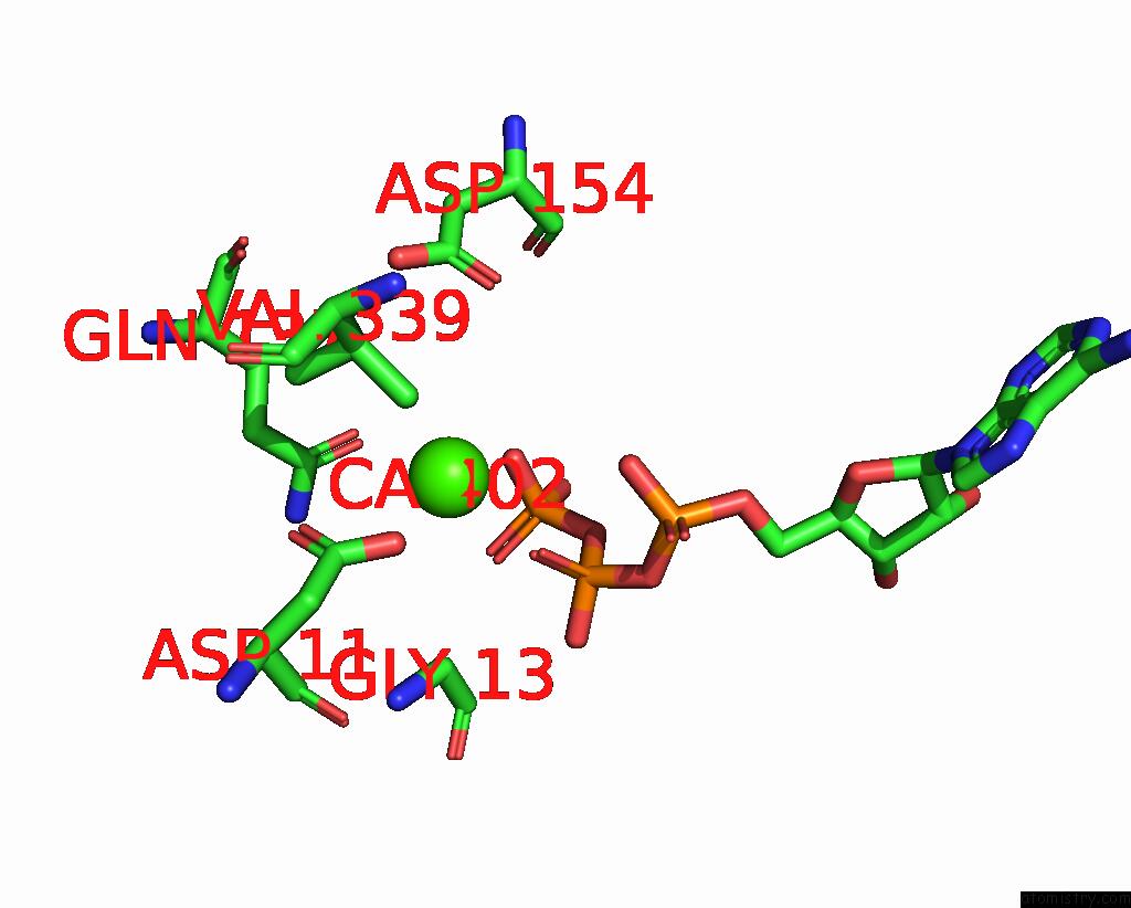
Calcium binding site 1 out of 12 in 4wyb

Go back to Calcium Binding Sites List in 4wyb
Calcium binding site 1 out of 12 in the Structure of the BUD6 Flank Domain in Complex with Actin


Mono view


Stereo pair view

A full contact list of Calcium with other atoms in the Ca binding site number 1 of Structure of the BUD6 Flank Domain in Complex with Actin within 5.0Å range:
probe atom residue distance (Å) B Occ
A:Ca402

b:62.2
occ:1.00
OE1 A:GLN137 2.5 69.6 1.0
O1B A:ATP401 2.5 72.3 1.0
O3G A:ATP401 2.7 74.2 1.0
O1G A:ATP401 3.7 67.7 1.0
PG A:ATP401 3.7 68.8 1.0
CD A:GLN137 3.7 63.9 1.0
OD2 A:ASP11 3.9 57.6 1.0
PB A:ATP401 3.9 74.0 1.0
O1A A:ATP401 4.3 76.4 1.0
O3B A:ATP401 4.3 66.6 1.0
CA A:GLY13 4.4 65.3 1.0
OD1 A:ASP154 4.4 78.5 1.0
OD1 A:ASP11 4.4 61.4 1.0
NE2 A:GLN137 4.5 66.5 1.0
CG A:ASP11 4.6 60.0 1.0
CG1 A:VAL339 4.6 63.1 1.0
CG A:GLN137 4.7 58.1 1.0
OD2 A:ASP154 4.8 72.0 1.0
O3A A:ATP401 4.8 77.5 1.0
O2B A:ATP401 4.8 68.3 1.0
NZ A:LYS18 4.9 65.7 1.0
CG A:ASP154 5.0 75.1 1.0
CB A:GLN137 5.0 63.6 1.0

Calcium binding site 2 out of 12 in 4wyb

Go back to Calcium Binding Sites List in 4wyb
Calcium binding site 2 out of 12 in the Structure of the BUD6 Flank Domain in Complex with Actin


Mono view


Stereo pair view

A full contact list of Calcium with other atoms in the Ca binding site number 2 of Structure of the BUD6 Flank Domain in Complex with Actin within 5.0Å range:
probe atom residue distance (Å) B Occ
C:Ca402

b:65.7
occ:1.00
O1B C:ATP401 2.5 91.7 1.0
OE1 C:GLN137 2.5 79.2 1.0
O1G C:ATP401 3.1 80.8 1.0
PB C:ATP401 3.6 82.6 1.0
CD C:GLN137 3.7 70.8 1.0
O3B C:ATP401 3.9 76.6 1.0
O3A C:ATP401 4.0 72.5 1.0
PG C:ATP401 4.0 78.3 1.0
OD2 C:ASP11 4.1 69.8 1.0
OD1 C:ASP154 4.3 73.3 1.0
CG1 C:VAL339 4.3 76.8 1.0
OD2 C:ASP154 4.4 74.9 1.0
NZ C:LYS18 4.5 80.3 1.0
NE2 C:GLN137 4.6 72.3 1.0
CG C:GLN137 4.6 60.8 1.0
OD1 C:ASP11 4.6 72.2 1.0
CG C:ASP154 4.7 70.9 1.0
O3G C:ATP401 4.8 74.6 1.0
CG C:ASP11 4.8 70.0 1.0
O2B C:ATP401 4.9 74.8 1.0
CE C:LYS18 5.0 78.0 1.0
CB C:GLN137 5.0 60.8 1.0
CB C:VAL339 5.0 62.8 1.0

Calcium binding site 3 out of 12 in 4wyb

Go back to Calcium Binding Sites List in 4wyb
Calcium binding site 3 out of 12 in the Structure of the BUD6 Flank Domain in Complex with Actin


Mono view


Stereo pair view

A full contact list of Calcium with other atoms in the Ca binding site number 3 of Structure of the BUD6 Flank Domain in Complex with Actin within 5.0Å range:
probe atom residue distance (Å) B Occ
E:Ca402

b:73.4
occ:1.00
O1B E:ATP401 2.4 88.3 1.0
OE1 E:GLN137 2.5 95.1 1.0
O2G E:ATP401 3.0 91.9 1.0
PB E:ATP401 3.5 91.9 1.0
PG E:ATP401 3.7 85.9 1.0
CD E:GLN137 3.7 80.9 1.0
O1G E:ATP401 3.8 80.3 1.0
O3B E:ATP401 3.8 85.8 1.0
OD2 E:ASP11 4.1 75.0 1.0
O3A E:ATP401 4.1 87.5 1.0
OD1 E:ASP154 4.4 88.7 1.0
NE2 E:GLN137 4.4 83.6 1.0
OD1 E:ASP11 4.5 78.2 1.0
CG1 E:VAL339 4.6 85.8 1.0
CG E:ASP11 4.7 76.0 1.0
NZ E:LYS18 4.7 86.0 1.0
OD2 E:ASP154 4.8 74.4 1.0
CG E:GLN137 4.8 73.5 1.0
CA E:GLY13 4.8 78.1 1.0
O2B E:ATP401 4.8 84.6 1.0
CG E:ASP154 5.0 81.1 1.0

Calcium binding site 4 out of 12 in 4wyb

Go back to Calcium Binding Sites List in 4wyb
Calcium binding site 4 out of 12 in the Structure of the BUD6 Flank Domain in Complex with Actin


Mono view


Stereo pair view

A full contact list of Calcium with other atoms in the Ca binding site number 4 of Structure of the BUD6 Flank Domain in Complex with Actin within 5.0Å range:
probe atom residue distance (Å) B Occ
G:Ca402

b:80.1
occ:1.00
OE1 G:GLN137 2.4 0.1 1.0
O2B G:ATP401 2.5 82.8 1.0
O1G G:ATP401 2.6 88.7 1.0
O2G G:ATP401 3.5 79.2 1.0
PG G:ATP401 3.5 83.2 1.0
CD G:GLN137 3.6 93.1 1.0
PB G:ATP401 3.8 89.7 1.0
O3B G:ATP401 4.2 80.6 1.0
OD2 G:ASP11 4.3 83.8 1.0
NE2 G:GLN137 4.4 95.4 1.0
O2A G:ATP401 4.5 89.0 1.0
CA G:GLY13 4.5 77.5 1.0
OD1 G:ASP11 4.5 86.2 1.0
OD1 G:ASP154 4.5 94.8 1.0
CG G:GLN137 4.6 84.6 1.0
CG2 G:VAL339 4.6 88.1 1.0
OD2 G:ASP154 4.7 79.4 1.0
O1B G:ATP401 4.8 86.3 1.0
O3A G:ATP401 4.8 90.8 1.0
CG G:ASP11 4.8 83.9 1.0
O3G G:ATP401 4.9 75.6 1.0
CB G:GLN137 4.9 82.1 1.0
CG G:ASP154 5.0 84.0 1.0
CG1 G:VAL339 5.0 80.4 1.0

Calcium binding site 5 out of 12 in 4wyb

Go back to Calcium Binding Sites List in 4wyb
Calcium binding site 5 out of 12 in the Structure of the BUD6 Flank Domain in Complex with Actin


Mono view


Stereo pair view

A full contact list of Calcium with other atoms in the Ca binding site number 5 of Structure of the BUD6 Flank Domain in Complex with Actin within 5.0Å range:
probe atom residue distance (Å) B Occ
I:Ca402

b:72.7
occ:1.00
O2B I:ATP401 2.5 68.8 1.0
NE2 I:GLN137 2.5 64.8 1.0
O3G I:ATP401 3.1 75.8 1.0
OD2 I:ASP11 3.7 55.4 1.0
O2A I:ATP401 3.8 80.8 1.0
PB I:ATP401 3.8 75.5 1.0
CD I:GLN137 3.8 59.1 1.0
PG I:ATP401 3.9 67.5 1.0
O2G I:ATP401 3.9 62.9 1.0
O3B I:ATP401 4.1 67.6 1.0
OD1 I:ASP11 4.3 56.9 1.0
CA I:GLY13 4.4 63.5 1.0
CG I:ASP11 4.4 57.2 1.0
OD1 I:ASP154 4.5 83.6 1.0
CG1 I:VAL339 4.5 63.6 1.0
CG I:GLN137 4.7 56.0 1.0
OE1 I:GLN137 4.7 66.0 1.0
NZ I:LYS18 4.7 67.6 1.0
O3A I:ATP401 4.7 76.1 1.0
PA I:ATP401 4.8 85.3 1.0
OD2 I:ASP154 4.8 73.9 1.0
O1B I:ATP401 4.8 74.2 1.0
CE I:LYS18 4.9 66.7 1.0
CB I:GLN137 4.9 61.1 1.0

Calcium binding site 6 out of 12 in 4wyb

Go back to Calcium Binding Sites List in 4wyb
Calcium binding site 6 out of 12 in the Structure of the BUD6 Flank Domain in Complex with Actin


Mono view


Stereo pair view

A full contact list of Calcium with other atoms in the Ca binding site number 6 of Structure of the BUD6 Flank Domain in Complex with Actin within 5.0Å range:
probe atom residue distance (Å) B Occ
K:Ca402

b:83.8
occ:1.00
O1B K:ATP401 2.4 91.0 1.0
OE1 K:GLN137 2.4 84.4 1.0
O1G K:ATP401 3.0 83.5 1.0
O2G K:ATP401 3.3 90.8 1.0
CD K:GLN137 3.6 84.2 1.0
PG K:ATP401 3.6 86.3 1.0
PB K:ATP401 3.8 95.7 1.0
OD2 K:ASP11 4.0 75.5 1.0
O3B K:ATP401 4.2 85.6 1.0
OD1 K:ASP11 4.2 76.1 1.0
NE2 K:GLN137 4.3 84.2 1.0
CA K:GLY13 4.3 90.6 1.0
OD1 K:ASP154 4.5 86.2 1.0
CG K:ASP11 4.6 81.6 1.0
CG K:GLN137 4.6 87.2 1.0
O2B K:ATP401 4.6 89.0 1.0
O1A K:ATP401 4.7 84.7 1.0
O3A K:ATP401 4.8 92.6 1.0
CG1 K:VAL339 4.8 98.4 1.0
CB K:GLN137 4.9 87.8 1.0

Calcium binding site 7 out of 12 in 4wyb

Go back to Calcium Binding Sites List in 4wyb
Calcium binding site 7 out of 12 in the Structure of the BUD6 Flank Domain in Complex with Actin


Mono view


Stereo pair view

A full contact list of Calcium with other atoms in the Ca binding site number 7 of Structure of the BUD6 Flank Domain in Complex with Actin within 5.0Å range:
probe atom residue distance (Å) B Occ
M:Ca402

b:67.3
occ:1.00
O1B M:ATP401 2.4 91.5 1.0
NE2 M:GLN137 2.8 72.9 1.0
PB M:ATP401 3.4 93.9 1.0
O2G M:ATP401 3.4 94.3 1.0
O3B M:ATP401 3.4 79.7 1.0
O3G M:ATP401 3.6 75.2 1.0
PG M:ATP401 3.7 83.6 1.0
CD M:GLN137 3.7 75.4 1.0
O3A M:ATP401 4.0 81.5 1.0
OE1 M:GLN137 4.1 80.5 1.0
OD2 M:ASP11 4.3 66.9 1.0
OD1 M:ASP154 4.5 74.9 1.0
OD2 M:ASP154 4.6 78.3 1.0
CG1 M:VAL339 4.6 76.7 1.0
OD1 M:ASP11 4.6 65.4 1.0
O2B M:ATP401 4.7 78.2 1.0
CE M:LYS18 4.8 94.8 1.0
CA M:GLY13 4.8 74.1 1.0
CG M:GLN137 4.8 59.9 1.0
CG M:ASP154 4.9 74.6 1.0
CG M:ASP11 4.9 67.4 1.0

Calcium binding site 8 out of 12 in 4wyb

Go back to Calcium Binding Sites List in 4wyb
Calcium binding site 8 out of 12 in the Structure of the BUD6 Flank Domain in Complex with Actin


Mono view


Stereo pair view

A full contact list of Calcium with other atoms in the Ca binding site number 8 of Structure of the BUD6 Flank Domain in Complex with Actin within 5.0Å range:
probe atom residue distance (Å) B Occ
O:Ca402

b:73.9
occ:1.00
OE1 O:GLN137 2.5 84.3 1.0
O2B O:ATP401 2.7 91.5 1.0
O2G O:ATP401 2.7 93.0 1.0
CD O:GLN137 3.4 81.0 1.0
PG O:ATP401 3.7 85.3 1.0
OD2 O:ASP11 3.8 77.5 1.0
O3G O:ATP401 3.8 88.1 1.0
NE2 O:GLN137 3.9 83.0 1.0
PB O:ATP401 4.0 98.6 1.0
O1A O:ATP401 4.1 81.6 1.0
O3B O:ATP401 4.2 85.3 1.0
OD1 O:ASP11 4.3 81.8 1.0
CA O:GLY13 4.4 86.8 1.0
CG O:ASP11 4.5 83.3 1.0
CG O:GLN137 4.5 80.9 1.0
CG1 O:VAL339 4.6 0.0 1.0
OD1 O:ASP154 4.6 82.1 1.0
O1B O:ATP401 4.8 99.4 1.0

Calcium binding site 9 out of 12 in 4wyb

Go back to Calcium Binding Sites List in 4wyb
Calcium binding site 9 out of 12 in the Structure of the BUD6 Flank Domain in Complex with Actin


Mono view


Stereo pair view

A full contact list of Calcium with other atoms in the Ca binding site number 9 of Structure of the BUD6 Flank Domain in Complex with Actin within 5.0Å range:
probe atom residue distance (Å) B Occ
Q:Ca402

b:68.3
occ:1.00
O2B Q:ATP401 2.4 75.2 1.0
OE1 Q:GLN137 2.4 82.0 1.0
O2G Q:ATP401 2.7 77.9 1.0
CD Q:GLN137 3.7 80.5 1.0
PG Q:ATP401 3.7 75.4 1.0
PB Q:ATP401 3.8 81.8 1.0
OD2 Q:ASP11 3.9 87.3 1.0
O3G Q:ATP401 3.9 77.5 1.0
O1A Q:ATP401 4.2 80.3 1.0
O3B Q:ATP401 4.2 77.8 1.0
OD1 Q:ASP11 4.4 80.0 1.0
NE2 Q:GLN137 4.5 77.0 1.0
CA Q:GLY13 4.5 78.4 1.0
OD1 Q:ASP154 4.5 80.8 1.0
O1B Q:ATP401 4.6 86.5 1.0
CG Q:ASP11 4.6 82.1 1.0
CG1 Q:VAL339 4.6 93.7 1.0
OD2 Q:ASP154 4.7 81.0 1.0
CG Q:GLN137 4.7 75.4 1.0
O3A Q:ATP401 4.9 83.0 1.0
PA Q:ATP401 5.0 89.2 1.0
NZ Q:LYS18 5.0 96.5 1.0
CG Q:ASP154 5.0 77.6 1.0

Calcium binding site 10 out of 12 in 4wyb

Go back to Calcium Binding Sites List in 4wyb
Calcium binding site 10 out of 12 in the Structure of the BUD6 Flank Domain in Complex with Actin


Mono view


Stereo pair view

A full contact list of Calcium with other atoms in the Ca binding site number 10 of Structure of the BUD6 Flank Domain in Complex with Actin within 5.0Å range:
probe atom residue distance (Å) B Occ
S:Ca402

b:66.8
occ:1.00
OE1 S:GLN137 2.5 72.3 1.0
O2B S:ATP401 2.5 88.2 1.0
O2G S:ATP401 2.7 77.3 1.0
CD S:GLN137 3.6 69.1 1.0
PB S:ATP401 3.7 81.9 1.0
O3B S:ATP401 3.8 75.5 1.0
PG S:ATP401 3.8 77.6 1.0
OD2 S:ASP11 4.0 62.4 1.0
OD1 S:ASP154 4.2 80.9 1.0
O3A S:ATP401 4.3 77.4 1.0
CG1 S:VAL339 4.3 75.2 1.0
OD2 S:ASP154 4.3 72.6 1.0
NE2 S:GLN137 4.4 68.2 1.0
CG S:GLN137 4.5 62.5 1.0
O3G S:ATP401 4.6 72.9 1.0
CG S:ASP154 4.6 75.2 1.0
OD1 S:ASP11 4.7 72.2 1.0
NZ S:LYS18 4.8 79.4 1.0
CG S:ASP11 4.8 65.9 1.0
O1B S:ATP401 4.9 80.9 1.0
O2A S:ATP401 4.9 78.7 1.0
CA S:GLY13 5.0 69.9 1.0
CG2 S:VAL339 5.0 63.3 1.0

Reference:

E.Park, B.R.Graziano, W.Zheng, M.Garabedian, B.L.Goode, M.J.Eck. Structure of A BUD6/Actin Complex Reveals A Novel WH2-Like Actin Monomer Recruitment Motif. Structure V. 23 1492 2015.
ISSN: ISSN 0969-2126
PubMed: 26118535
DOI: 10.1016/J.STR.2015.05.015
Page generated: Sun Jul 14 14:25:39 2024

Last articles

Zn in 9MJ5
Zn in 9HNW
Zn in 9G0L
Zn in 9FNE
Zn in 9DZN
Zn in 9E0I
Zn in 9D32
Zn in 9DAK
Zn in 8ZXC
Zn in 8ZUF
© Copyright 2008-2020 by atomistry.com
Home   |    Site Map   |    Copyright   |    Contact us   |    Privacy