Calcium »
PDB 4x8e-4xur »
4xb3 »
Calcium in PDB 4xb3: Structure of Dextran Glucosidase
Enzymatic activity of Structure of Dextran Glucosidase
All present enzymatic activity of Structure of Dextran Glucosidase:
3.2.1.70;
3.2.1.70;
Protein crystallography data
The structure of Structure of Dextran Glucosidase, PDB code: 4xb3
was solved by
M.Kobayashi,
K.Kato,
M.Yao,
with X-Ray Crystallography technique. A brief refinement statistics is given in the table below:
| Resolution Low / High (Å) | 48.11 / 2.09 |
| Space group | P 21 21 21 |
| Cell size a, b, c (Å), α, β, γ (°) | 72.151, 82.550, 103.559, 90.00, 90.00, 90.00 |
| R / Rfree (%) | 17.9 / 21.9 |
Calcium Binding Sites:
The binding sites of Calcium atom in the Structure of Dextran Glucosidase
(pdb code 4xb3). This binding sites where shown within
5.0 Angstroms radius around Calcium atom.
In total 4 binding sites of Calcium where determined in the Structure of Dextran Glucosidase, PDB code: 4xb3:
Jump to Calcium binding site number: 1; 2; 3; 4;
In total 4 binding sites of Calcium where determined in the Structure of Dextran Glucosidase, PDB code: 4xb3:
Jump to Calcium binding site number: 1; 2; 3; 4;
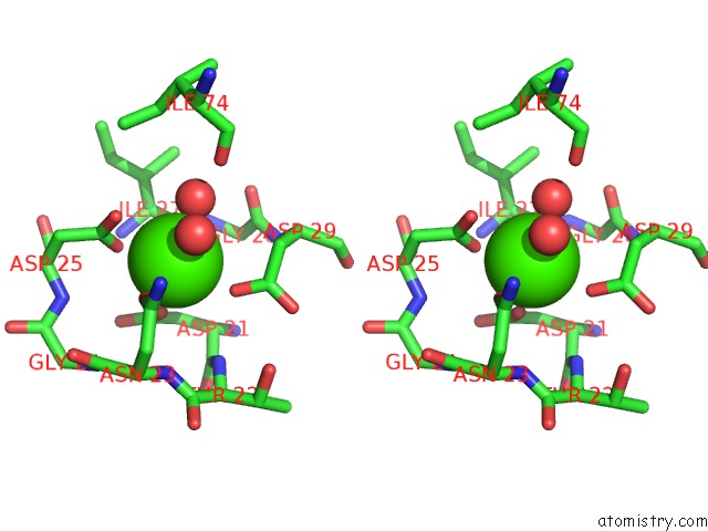
Calcium binding site 1 out of 4 in 4xb3
Go back to
Calcium binding site 1 out
of 4 in the Structure of Dextran Glucosidase

Mono view

Stereo pair view
Mono view
Stereo pair view
A full contact list of Calcium with other atoms in the Ca binding
site number 1 of Structure of Dextran Glucosidase within 5.0Å range:
|
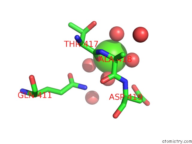
Calcium binding site 2 out of 4 in 4xb3
Go back to
Calcium binding site 2 out
of 4 in the Structure of Dextran Glucosidase

Mono view
Stereo pair view
Mono view
Stereo pair view
A full contact list of Calcium with other atoms in the Ca binding
site number 2 of Structure of Dextran Glucosidase within 5.0Å range:
|
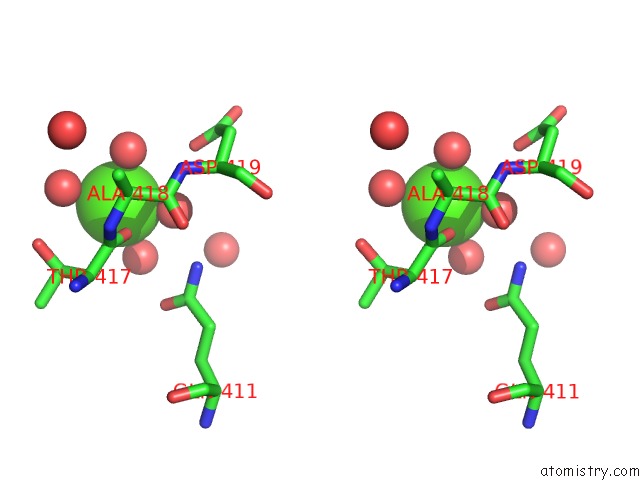
Calcium binding site 3 out of 4 in 4xb3
Go back to
Calcium binding site 3 out
of 4 in the Structure of Dextran Glucosidase
Mono view
Stereo pair view
Mono view
Stereo pair view
A full contact list of Calcium with other atoms in the Ca binding
site number 3 of Structure of Dextran Glucosidase within 5.0Å range:
|
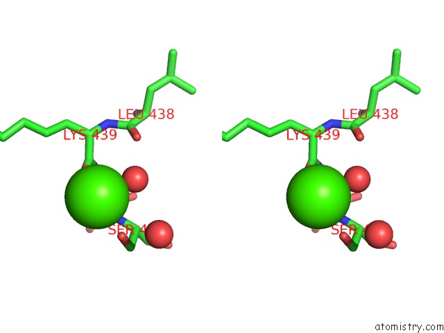
Calcium binding site 4 out of 4 in 4xb3
Go back to
Calcium binding site 4 out
of 4 in the Structure of Dextran Glucosidase

Mono view
Stereo pair view
Mono view
Stereo pair view
A full contact list of Calcium with other atoms in the Ca binding
site number 4 of Structure of Dextran Glucosidase within 5.0Å range:
|
Reference:
M.Kobayashi,
W.Saburi,
D.Nakatsuka,
H.Hondoh,
K.Kato,
M.Okuyama,
H.Mori,
A.Kimura,
M.Yao.
Structural Insights Into the Catalytic Reaction That Is Involved in the Reorientation of TRP238 at the Substrate-Binding Site in GH13 Dextran Glucosidase Febs Lett. V. 589 484 2015.
ISSN: ISSN 0014-5793
PubMed: 25595454
DOI: 10.1016/J.FEBSLET.2015.01.005
Page generated: Wed Jul 9 02:51:15 2025
ISSN: ISSN 0014-5793
PubMed: 25595454
DOI: 10.1016/J.FEBSLET.2015.01.005
Last articles
I in 3UWPI in 3USM
I in 3USL
I in 3TSG
I in 3UNH
I in 3UNF
I in 3UBA
I in 3UFM
I in 3TUR
I in 3UCO