Calcium »
PDB 4xut-4y9b »
4xym »
Calcium in PDB 4xym: Ca. Korarchaeum Cryptofilum Dinucleotide Forming Acetyl-Coenzyme A Synthetase 1 in Complex with Coenzyme A, Ca-Ampcp and Hgcl+
Protein crystallography data
The structure of Ca. Korarchaeum Cryptofilum Dinucleotide Forming Acetyl-Coenzyme A Synthetase 1 in Complex with Coenzyme A, Ca-Ampcp and Hgcl+, PDB code: 4xym
was solved by
R.H.-J.Weisse,
A.J.Scheidig,
with X-Ray Crystallography technique. A brief refinement statistics is given in the table below:
| Resolution Low / High (Å) | 85.02 / 1.90 |
| Space group | P 21 21 21 |
| Cell size a, b, c (Å), α, β, γ (°) | 99.560, 114.416, 127.040, 90.00, 90.00, 90.00 |
| R / Rfree (%) | 19.5 / 23.7 |
Other elements in 4xym:
The structure of Ca. Korarchaeum Cryptofilum Dinucleotide Forming Acetyl-Coenzyme A Synthetase 1 in Complex with Coenzyme A, Ca-Ampcp and Hgcl+ also contains other interesting chemical elements:
| Mercury | (Hg) | 8 atoms |
| Chlorine | (Cl) | 6 atoms |
| Sodium | (Na) | 1 atom |
Calcium Binding Sites:
The binding sites of Calcium atom in the Ca. Korarchaeum Cryptofilum Dinucleotide Forming Acetyl-Coenzyme A Synthetase 1 in Complex with Coenzyme A, Ca-Ampcp and Hgcl+
(pdb code 4xym). This binding sites where shown within
5.0 Angstroms radius around Calcium atom.
In total 2 binding sites of Calcium where determined in the Ca. Korarchaeum Cryptofilum Dinucleotide Forming Acetyl-Coenzyme A Synthetase 1 in Complex with Coenzyme A, Ca-Ampcp and Hgcl+, PDB code: 4xym:
Jump to Calcium binding site number: 1; 2;
In total 2 binding sites of Calcium where determined in the Ca. Korarchaeum Cryptofilum Dinucleotide Forming Acetyl-Coenzyme A Synthetase 1 in Complex with Coenzyme A, Ca-Ampcp and Hgcl+, PDB code: 4xym:
Jump to Calcium binding site number: 1; 2;
Calcium binding site 1 out of 2 in 4xym
Go back to
Calcium binding site 1 out
of 2 in the Ca. Korarchaeum Cryptofilum Dinucleotide Forming Acetyl-Coenzyme A Synthetase 1 in Complex with Coenzyme A, Ca-Ampcp and Hgcl+
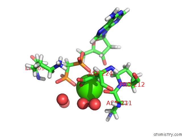
Mono view
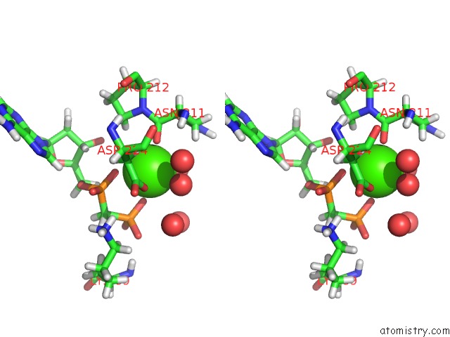
Stereo pair view
Mono view
Stereo pair view
A full contact list of Calcium with other atoms in the Ca binding
site number 1 of Ca. Korarchaeum Cryptofilum Dinucleotide Forming Acetyl-Coenzyme A Synthetase 1 in Complex with Coenzyme A, Ca-Ampcp and Hgcl+ within 5.0Å range:
|
Calcium binding site 2 out of 2 in 4xym
Go back to
Calcium binding site 2 out
of 2 in the Ca. Korarchaeum Cryptofilum Dinucleotide Forming Acetyl-Coenzyme A Synthetase 1 in Complex with Coenzyme A, Ca-Ampcp and Hgcl+
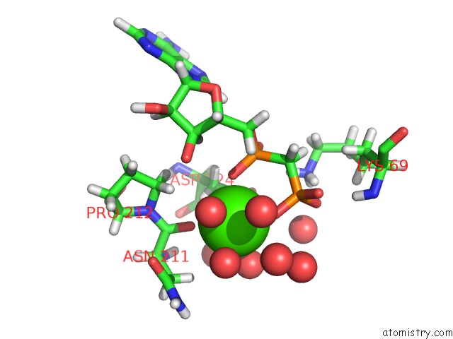
Mono view
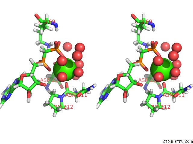
Stereo pair view
Mono view
Stereo pair view
A full contact list of Calcium with other atoms in the Ca binding
site number 2 of Ca. Korarchaeum Cryptofilum Dinucleotide Forming Acetyl-Coenzyme A Synthetase 1 in Complex with Coenzyme A, Ca-Ampcp and Hgcl+ within 5.0Å range:
|
Reference:
R.H.Weie,
A.Faust,
M.Schmidt,
P.Schonheit,
A.J.Scheidig.
Structure of Ndp-Forming Acetyl-Coa Synthetase ACD1 Reveals A Large Rearrangement For Phosphoryl Transfer. Proc.Natl.Acad.Sci.Usa V. 113 E519 2016.
ISSN: ESSN 1091-6490
PubMed: 26787904
DOI: 10.1073/PNAS.1518614113
Page generated: Wed Jul 9 02:59:23 2025
ISSN: ESSN 1091-6490
PubMed: 26787904
DOI: 10.1073/PNAS.1518614113
Last articles
Mn in 9LJUMn in 9LJW
Mn in 9LJS
Mn in 9LJR
Mn in 9LJT
Mn in 9LJV
Mg in 9UA2
Mg in 9R96
Mg in 9VM1
Mg in 9P01