Calcium »
PDB 4xut-4y9b »
4y1t »
Calcium in PDB 4y1t: Structural Basis For CA2+-Mediated Interaction of the Perforin C2 Domain with Lipid Membranes
Protein crystallography data
The structure of Structural Basis For CA2+-Mediated Interaction of the Perforin C2 Domain with Lipid Membranes, PDB code: 4y1t
was solved by
P.J.Conroy,
H.Yagi,
J.C.Whisstock,
R.S.Norton,
with X-Ray Crystallography technique. A brief refinement statistics is given in the table below:
| Resolution Low / High (Å) | 42.38 / 2.67 |
| Space group | I 2 2 2 |
| Cell size a, b, c (Å), α, β, γ (°) | 45.009, 59.828, 125.919, 90.00, 90.00, 90.00 |
| R / Rfree (%) | 16.8 / 21.5 |
Calcium Binding Sites:
The binding sites of Calcium atom in the Structural Basis For CA2+-Mediated Interaction of the Perforin C2 Domain with Lipid Membranes
(pdb code 4y1t). This binding sites where shown within
5.0 Angstroms radius around Calcium atom.
In total 5 binding sites of Calcium where determined in the Structural Basis For CA2+-Mediated Interaction of the Perforin C2 Domain with Lipid Membranes, PDB code: 4y1t:
Jump to Calcium binding site number: 1; 2; 3; 4; 5;
In total 5 binding sites of Calcium where determined in the Structural Basis For CA2+-Mediated Interaction of the Perforin C2 Domain with Lipid Membranes, PDB code: 4y1t:
Jump to Calcium binding site number: 1; 2; 3; 4; 5;
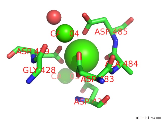
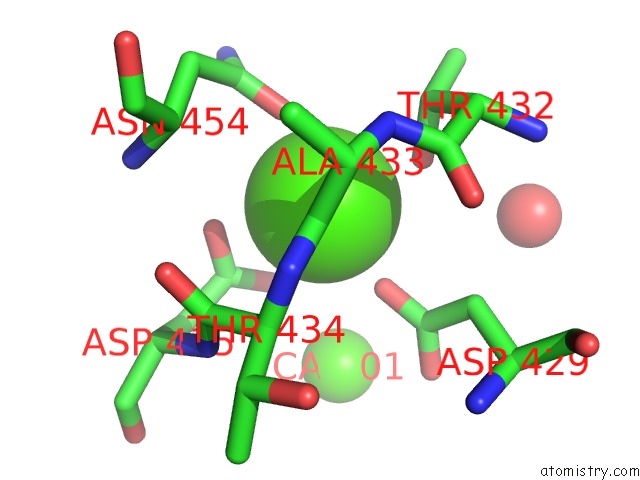
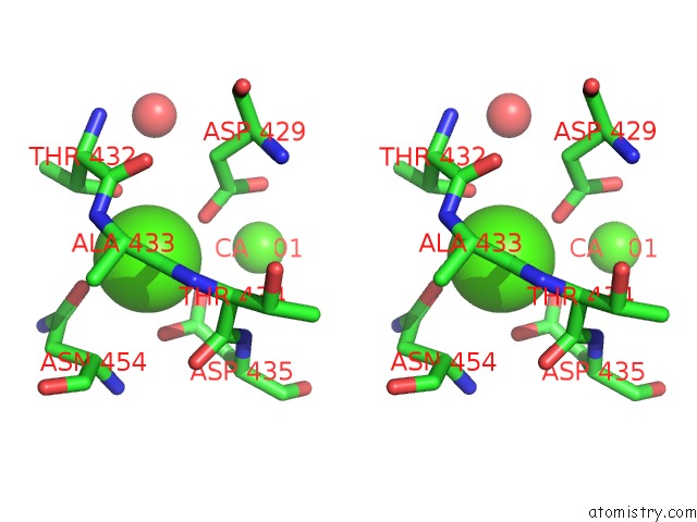
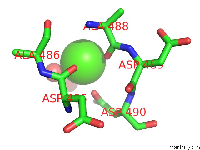
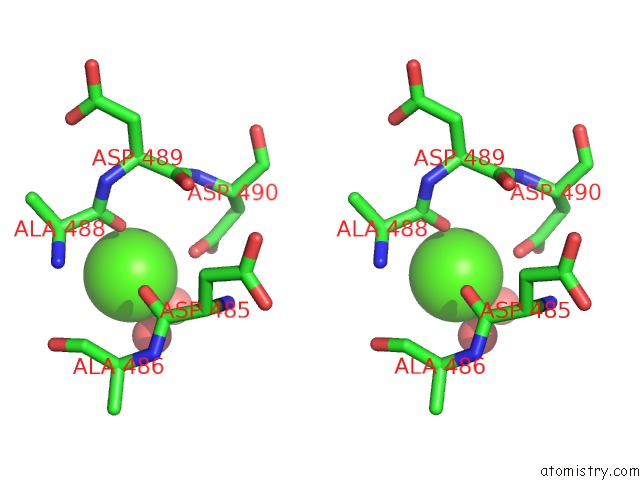
Calcium binding site 1 out of 5 in 4y1t
Go back to
Calcium binding site 1 out
of 5 in the Structural Basis For CA2+-Mediated Interaction of the Perforin C2 Domain with Lipid Membranes
Mono view
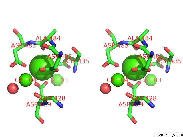
Stereo pair view
Mono view
Stereo pair view
A full contact list of Calcium with other atoms in the Ca binding
site number 1 of Structural Basis For CA2+-Mediated Interaction of the Perforin C2 Domain with Lipid Membranes within 5.0Å range:
|
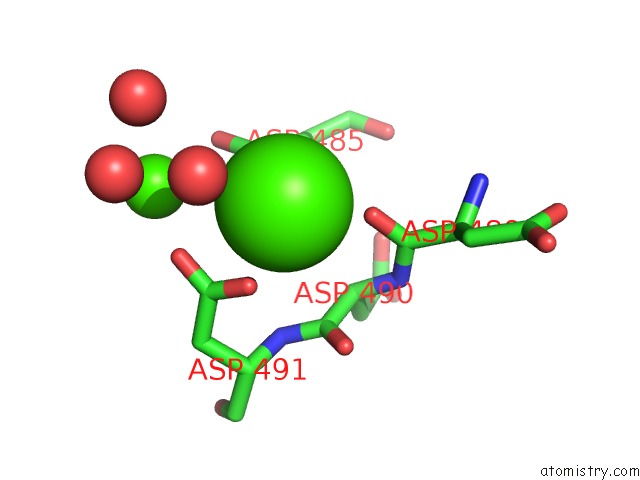
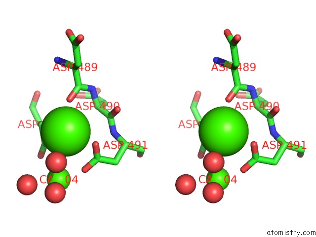
Calcium binding site 2 out of 5 in 4y1t
Go back to
Calcium binding site 2 out
of 5 in the Structural Basis For CA2+-Mediated Interaction of the Perforin C2 Domain with Lipid Membranes
Mono view
Stereo pair view
Mono view
Stereo pair view
A full contact list of Calcium with other atoms in the Ca binding
site number 2 of Structural Basis For CA2+-Mediated Interaction of the Perforin C2 Domain with Lipid Membranes within 5.0Å range:
|
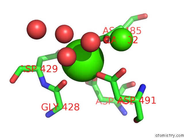
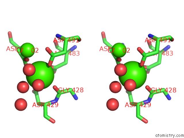
Calcium binding site 3 out of 5 in 4y1t
Go back to
Calcium binding site 3 out
of 5 in the Structural Basis For CA2+-Mediated Interaction of the Perforin C2 Domain with Lipid Membranes
Mono view
Stereo pair view
Mono view
Stereo pair view
A full contact list of Calcium with other atoms in the Ca binding
site number 3 of Structural Basis For CA2+-Mediated Interaction of the Perforin C2 Domain with Lipid Membranes within 5.0Å range:
|
Calcium binding site 4 out of 5 in 4y1t
Go back to
Calcium binding site 4 out
of 5 in the Structural Basis For CA2+-Mediated Interaction of the Perforin C2 Domain with Lipid Membranes
Mono view
Stereo pair view
Mono view
Stereo pair view
A full contact list of Calcium with other atoms in the Ca binding
site number 4 of Structural Basis For CA2+-Mediated Interaction of the Perforin C2 Domain with Lipid Membranes within 5.0Å range:
|
Calcium binding site 5 out of 5 in 4y1t
Go back to
Calcium binding site 5 out
of 5 in the Structural Basis For CA2+-Mediated Interaction of the Perforin C2 Domain with Lipid Membranes
Mono view
Stereo pair view
Mono view
Stereo pair view
A full contact list of Calcium with other atoms in the Ca binding
site number 5 of Structural Basis For CA2+-Mediated Interaction of the Perforin C2 Domain with Lipid Membranes within 5.0Å range:
|
Reference:
H.Yagi,
P.J.Conroy,
E.W.Leung,
R.H.Law,
J.A.Trapani,
I.Voskoboinik,
J.C.Whisstock,
R.S.Norton.
Structural Basis For CA2+-Mediated Interaction of the Perforin C2 Domain with Lipid Membranes. J.Biol.Chem. V. 290 25213 2015.
ISSN: ESSN 1083-351X
PubMed: 26306037
DOI: 10.1074/JBC.M115.668384
Page generated: Wed Jul 9 03:01:12 2025
ISSN: ESSN 1083-351X
PubMed: 26306037
DOI: 10.1074/JBC.M115.668384
Last articles
Cl in 5ZZNCl in 6A15
Cl in 5ZWY
Cl in 6A02
Cl in 5ZZG
Cl in 5ZXF
Cl in 5ZZC
Cl in 5ZXE
Cl in 5ZXL
Cl in 5ZW8