Calcium »
PDB 5a4x-5afp »
5a6x »
Calcium in PDB 5a6x: Structure of the Lecb Lectin From Pseudomonas Aeruginosa Strain PA14 in Complex with Alpha-Methyl-Fucoside
Protein crystallography data
The structure of Structure of the Lecb Lectin From Pseudomonas Aeruginosa Strain PA14 in Complex with Alpha-Methyl-Fucoside, PDB code: 5a6x
was solved by
R.Sommer,
S.Wagner,
A.Varrot,
A.Khaledi,
S.Haussler,
A.Imberty,
A.Titz,
with X-Ray Crystallography technique. A brief refinement statistics is given in the table below:
| Resolution Low / High (Å) | 37.84 / 1.55 |
| Space group | P 21 21 21 |
| Cell size a, b, c (Å), α, β, γ (°) | 52.558, 65.585, 109.036, 90.00, 90.00, 90.00 |
| R / Rfree (%) | 12.8 / 15.3 |
Calcium Binding Sites:
The binding sites of Calcium atom in the Structure of the Lecb Lectin From Pseudomonas Aeruginosa Strain PA14 in Complex with Alpha-Methyl-Fucoside
(pdb code 5a6x). This binding sites where shown within
5.0 Angstroms radius around Calcium atom.
In total 8 binding sites of Calcium where determined in the Structure of the Lecb Lectin From Pseudomonas Aeruginosa Strain PA14 in Complex with Alpha-Methyl-Fucoside, PDB code: 5a6x:
Jump to Calcium binding site number: 1; 2; 3; 4; 5; 6; 7; 8;
In total 8 binding sites of Calcium where determined in the Structure of the Lecb Lectin From Pseudomonas Aeruginosa Strain PA14 in Complex with Alpha-Methyl-Fucoside, PDB code: 5a6x:
Jump to Calcium binding site number: 1; 2; 3; 4; 5; 6; 7; 8;
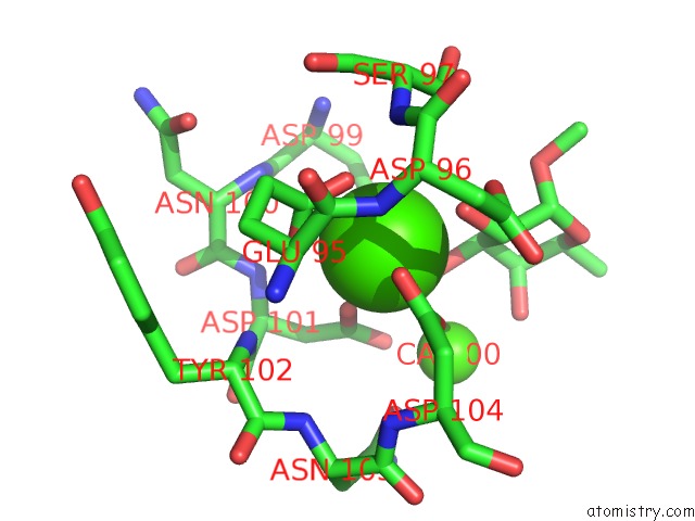
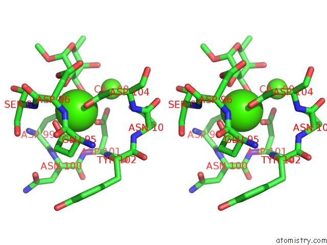
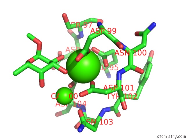
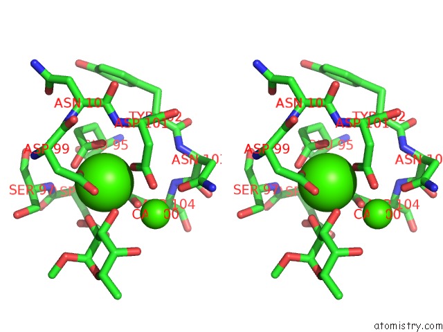
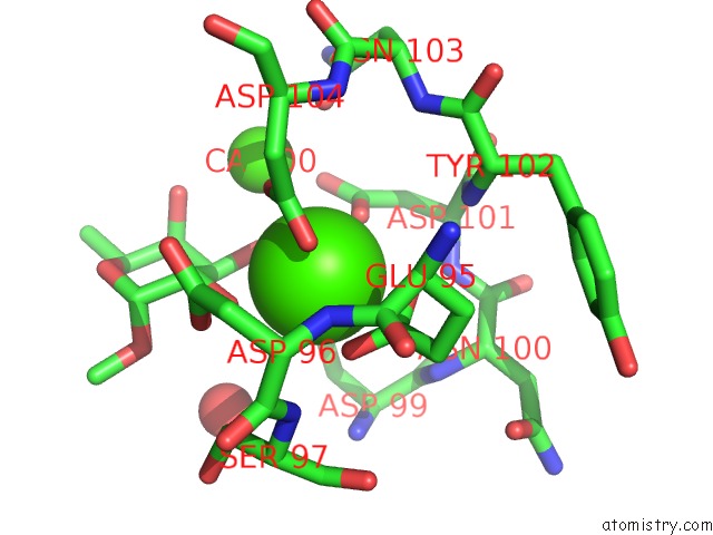
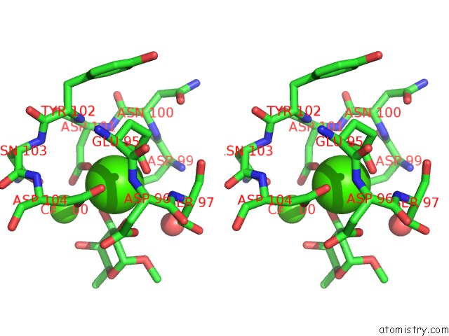
Calcium binding site 1 out of 8 in 5a6x
Go back to
Calcium binding site 1 out
of 8 in the Structure of the Lecb Lectin From Pseudomonas Aeruginosa Strain PA14 in Complex with Alpha-Methyl-Fucoside
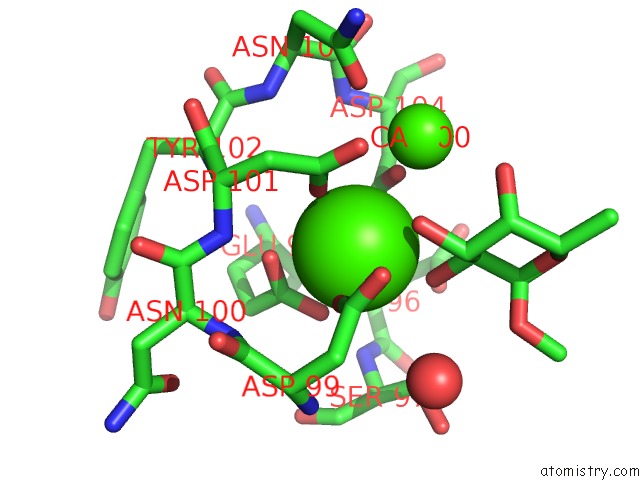
Mono view
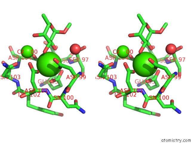
Stereo pair view
Mono view
Stereo pair view
A full contact list of Calcium with other atoms in the Ca binding
site number 1 of Structure of the Lecb Lectin From Pseudomonas Aeruginosa Strain PA14 in Complex with Alpha-Methyl-Fucoside within 5.0Å range:
|
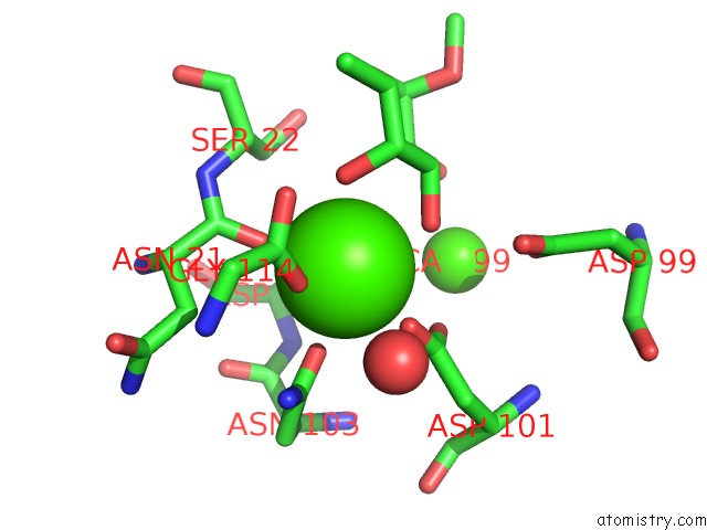
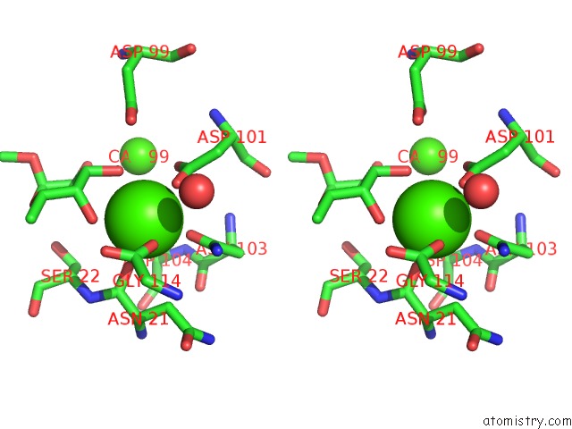
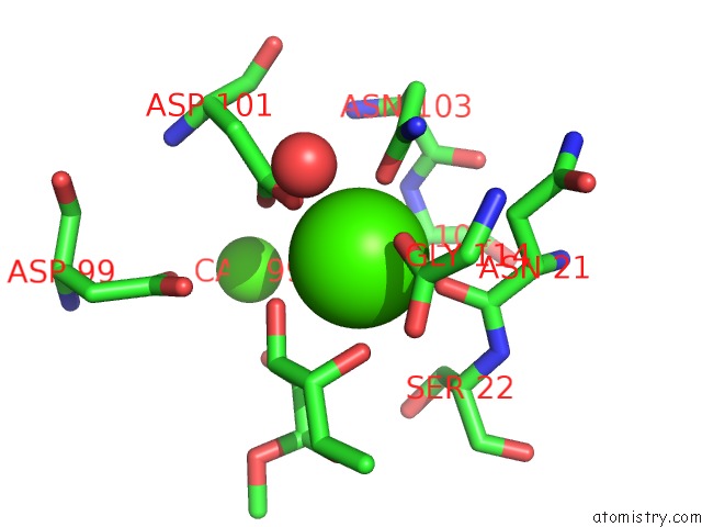
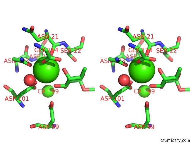
Calcium binding site 2 out of 8 in 5a6x
Go back to
Calcium binding site 2 out
of 8 in the Structure of the Lecb Lectin From Pseudomonas Aeruginosa Strain PA14 in Complex with Alpha-Methyl-Fucoside
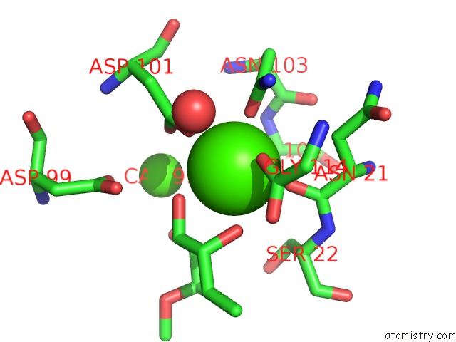
Mono view
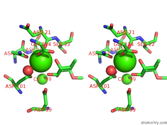
Stereo pair view
Mono view
Stereo pair view
A full contact list of Calcium with other atoms in the Ca binding
site number 2 of Structure of the Lecb Lectin From Pseudomonas Aeruginosa Strain PA14 in Complex with Alpha-Methyl-Fucoside within 5.0Å range:
|
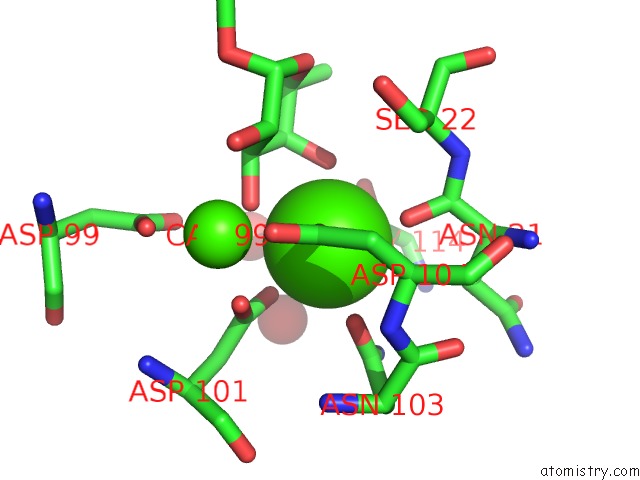
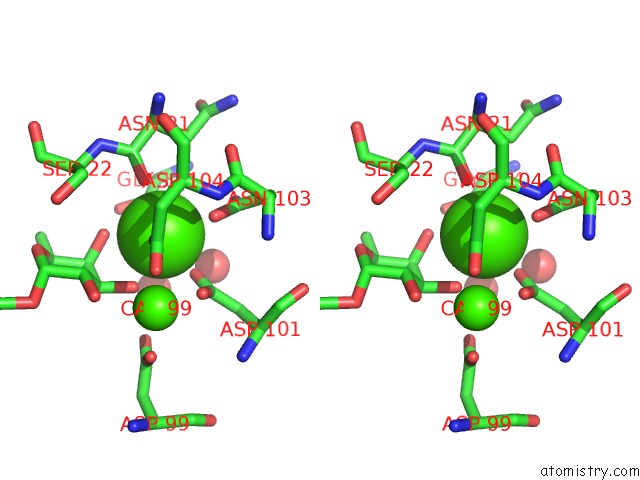
Calcium binding site 3 out of 8 in 5a6x
Go back to
Calcium binding site 3 out
of 8 in the Structure of the Lecb Lectin From Pseudomonas Aeruginosa Strain PA14 in Complex with Alpha-Methyl-Fucoside
Mono view
Stereo pair view
Mono view
Stereo pair view
A full contact list of Calcium with other atoms in the Ca binding
site number 3 of Structure of the Lecb Lectin From Pseudomonas Aeruginosa Strain PA14 in Complex with Alpha-Methyl-Fucoside within 5.0Å range:
|
Calcium binding site 4 out of 8 in 5a6x
Go back to
Calcium binding site 4 out
of 8 in the Structure of the Lecb Lectin From Pseudomonas Aeruginosa Strain PA14 in Complex with Alpha-Methyl-Fucoside
Mono view
Stereo pair view
Mono view
Stereo pair view
A full contact list of Calcium with other atoms in the Ca binding
site number 4 of Structure of the Lecb Lectin From Pseudomonas Aeruginosa Strain PA14 in Complex with Alpha-Methyl-Fucoside within 5.0Å range:
|
Calcium binding site 5 out of 8 in 5a6x
Go back to
Calcium binding site 5 out
of 8 in the Structure of the Lecb Lectin From Pseudomonas Aeruginosa Strain PA14 in Complex with Alpha-Methyl-Fucoside
Mono view
Stereo pair view
Mono view
Stereo pair view
A full contact list of Calcium with other atoms in the Ca binding
site number 5 of Structure of the Lecb Lectin From Pseudomonas Aeruginosa Strain PA14 in Complex with Alpha-Methyl-Fucoside within 5.0Å range:
|
Calcium binding site 6 out of 8 in 5a6x
Go back to
Calcium binding site 6 out
of 8 in the Structure of the Lecb Lectin From Pseudomonas Aeruginosa Strain PA14 in Complex with Alpha-Methyl-Fucoside
Mono view
Stereo pair view
Mono view
Stereo pair view
A full contact list of Calcium with other atoms in the Ca binding
site number 6 of Structure of the Lecb Lectin From Pseudomonas Aeruginosa Strain PA14 in Complex with Alpha-Methyl-Fucoside within 5.0Å range:
|
Calcium binding site 7 out of 8 in 5a6x
Go back to
Calcium binding site 7 out
of 8 in the Structure of the Lecb Lectin From Pseudomonas Aeruginosa Strain PA14 in Complex with Alpha-Methyl-Fucoside
Mono view
Stereo pair view
Mono view
Stereo pair view
A full contact list of Calcium with other atoms in the Ca binding
site number 7 of Structure of the Lecb Lectin From Pseudomonas Aeruginosa Strain PA14 in Complex with Alpha-Methyl-Fucoside within 5.0Å range:
|
Calcium binding site 8 out of 8 in 5a6x
Go back to
Calcium binding site 8 out
of 8 in the Structure of the Lecb Lectin From Pseudomonas Aeruginosa Strain PA14 in Complex with Alpha-Methyl-Fucoside
Mono view
Stereo pair view
Mono view
Stereo pair view
A full contact list of Calcium with other atoms in the Ca binding
site number 8 of Structure of the Lecb Lectin From Pseudomonas Aeruginosa Strain PA14 in Complex with Alpha-Methyl-Fucoside within 5.0Å range:
|
Reference:
R.Sommer,
S.Wagner,
A.Varrot,
C.M.Nycholat,
A.Khaledi,
S.Haussler,
J.C.Paulson,
A.Imberty,
A.Titz.
The Virulence Factor Lecb Varies in Clinical Isolates: Consequences For Ligand Binding and Drug Discovery. Chem Sci V. 7 4990 2016.
ISSN: ISSN 2041-6520
PubMed: 30155149
DOI: 10.1039/C6SC00696E
Page generated: Wed Jul 9 04:04:14 2025
ISSN: ISSN 2041-6520
PubMed: 30155149
DOI: 10.1039/C6SC00696E
Last articles
Mg in 4KMUMg in 4KLN
Mg in 4KMR
Mg in 4KLO
Mg in 4KLZ
Mg in 4KLM
Mg in 4KLL
Mg in 4KLJ
Mg in 4KLI
Mg in 4KLG