Calcium »
PDB 5b5e-5byn »
5bmv »
Calcium in PDB 5bmv: Crystal Structure of Tubulin-Stathmin-Ttl-Vinblastine Complex
Protein crystallography data
The structure of Crystal Structure of Tubulin-Stathmin-Ttl-Vinblastine Complex, PDB code: 5bmv
was solved by
Y.Wang,
Q.Chen,
R.Zhang,
with X-Ray Crystallography technique. A brief refinement statistics is given in the table below:
| Resolution Low / High (Å) | 50.00 / 2.50 |
| Space group | P 21 21 21 |
| Cell size a, b, c (Å), α, β, γ (°) | 105.470, 157.420, 183.180, 90.00, 90.00, 90.00 |
| R / Rfree (%) | 18.4 / 23.5 |
Other elements in 5bmv:
The structure of Crystal Structure of Tubulin-Stathmin-Ttl-Vinblastine Complex also contains other interesting chemical elements:
| Magnesium | (Mg) | 4 atoms |
Calcium Binding Sites:
The binding sites of Calcium atom in the Crystal Structure of Tubulin-Stathmin-Ttl-Vinblastine Complex
(pdb code 5bmv). This binding sites where shown within
5.0 Angstroms radius around Calcium atom.
In total 3 binding sites of Calcium where determined in the Crystal Structure of Tubulin-Stathmin-Ttl-Vinblastine Complex, PDB code: 5bmv:
Jump to Calcium binding site number: 1; 2; 3;
In total 3 binding sites of Calcium where determined in the Crystal Structure of Tubulin-Stathmin-Ttl-Vinblastine Complex, PDB code: 5bmv:
Jump to Calcium binding site number: 1; 2; 3;
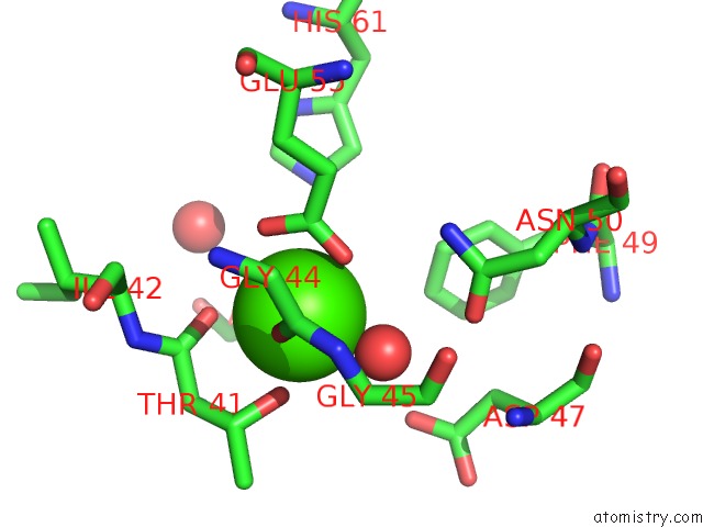
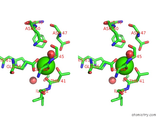
Calcium binding site 1 out of 3 in 5bmv
Go back to
Calcium binding site 1 out
of 3 in the Crystal Structure of Tubulin-Stathmin-Ttl-Vinblastine Complex
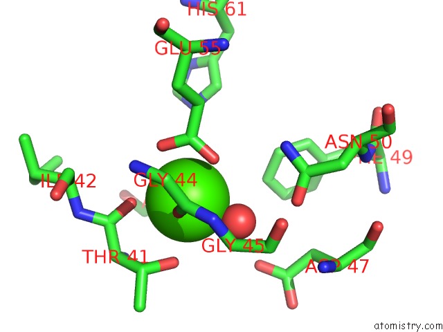
Mono view
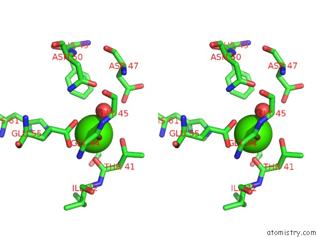
Stereo pair view
Mono view
Stereo pair view
A full contact list of Calcium with other atoms in the Ca binding
site number 1 of Crystal Structure of Tubulin-Stathmin-Ttl-Vinblastine Complex within 5.0Å range:
|
Calcium binding site 2 out of 3 in 5bmv
Go back to
Calcium binding site 2 out
of 3 in the Crystal Structure of Tubulin-Stathmin-Ttl-Vinblastine Complex
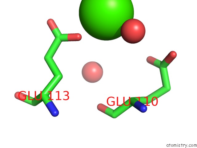
Mono view
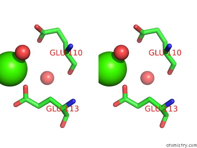
Stereo pair view
Mono view
Stereo pair view
A full contact list of Calcium with other atoms in the Ca binding
site number 2 of Crystal Structure of Tubulin-Stathmin-Ttl-Vinblastine Complex within 5.0Å range:
|
Calcium binding site 3 out of 3 in 5bmv
Go back to
Calcium binding site 3 out
of 3 in the Crystal Structure of Tubulin-Stathmin-Ttl-Vinblastine Complex
Mono view
Stereo pair view
Mono view
Stereo pair view
A full contact list of Calcium with other atoms in the Ca binding
site number 3 of Crystal Structure of Tubulin-Stathmin-Ttl-Vinblastine Complex within 5.0Å range:
|
Reference:
Y.Wang,
F.W.Benz,
Y.Wu,
Q.Wang,
Y.Chen,
X.Chen,
H.Li,
Y.Zhang,
R.Zhang,
J.Yang.
Structural Insights Into the Pharmacophore of Vinca Domain Inhibitors of Microtubules. Mol.Pharmacol. V. 89 233 2016.
ISSN: ESSN 1521-0111
PubMed: 26660762
DOI: 10.1124/MOL.115.100149
Page generated: Wed Jul 9 04:25:47 2025
ISSN: ESSN 1521-0111
PubMed: 26660762
DOI: 10.1124/MOL.115.100149
Last articles
Mg in 7BGIMg in 7BLX
Mg in 7BLZ
Mg in 7BOD
Mg in 7BNR
Mg in 7BNK
Mg in 7BMC
Mg in 7BM9
Mg in 7BM8
Mg in 7BM6