Calcium »
PDB 5b5e-5byn »
5bvu »
Calcium in PDB 5bvu: Crystal Structure of Thermoanaerobacterium Xylolyticum GH116 Beta- Glucosidase
Enzymatic activity of Crystal Structure of Thermoanaerobacterium Xylolyticum GH116 Beta- Glucosidase
All present enzymatic activity of Crystal Structure of Thermoanaerobacterium Xylolyticum GH116 Beta- Glucosidase:
3.2.1.21;
3.2.1.21;
Protein crystallography data
The structure of Crystal Structure of Thermoanaerobacterium Xylolyticum GH116 Beta- Glucosidase, PDB code: 5bvu
was solved by
R.Charoenwattanasatien,
S.Pengthaisong,
S.Sansenya,
R.Mutoh,
H.Tanaka,
G.Kurisu,
J.R.Ketudat Cairns,
with X-Ray Crystallography technique. A brief refinement statistics is given in the table below:
| Resolution Low / High (Å) | 50.00 / 1.61 |
| Space group | P 21 21 2 |
| Cell size a, b, c (Å), α, β, γ (°) | 177.683, 54.271, 83.180, 90.00, 90.00, 90.00 |
| R / Rfree (%) | 15.4 / 17.5 |
Calcium Binding Sites:
The binding sites of Calcium atom in the Crystal Structure of Thermoanaerobacterium Xylolyticum GH116 Beta- Glucosidase
(pdb code 5bvu). This binding sites where shown within
5.0 Angstroms radius around Calcium atom.
In total only one binding site of Calcium was determined in the Crystal Structure of Thermoanaerobacterium Xylolyticum GH116 Beta- Glucosidase, PDB code: 5bvu:
In total only one binding site of Calcium was determined in the Crystal Structure of Thermoanaerobacterium Xylolyticum GH116 Beta- Glucosidase, PDB code: 5bvu:
Calcium binding site 1 out of 1 in 5bvu
Go back to
Calcium binding site 1 out
of 1 in the Crystal Structure of Thermoanaerobacterium Xylolyticum GH116 Beta- Glucosidase
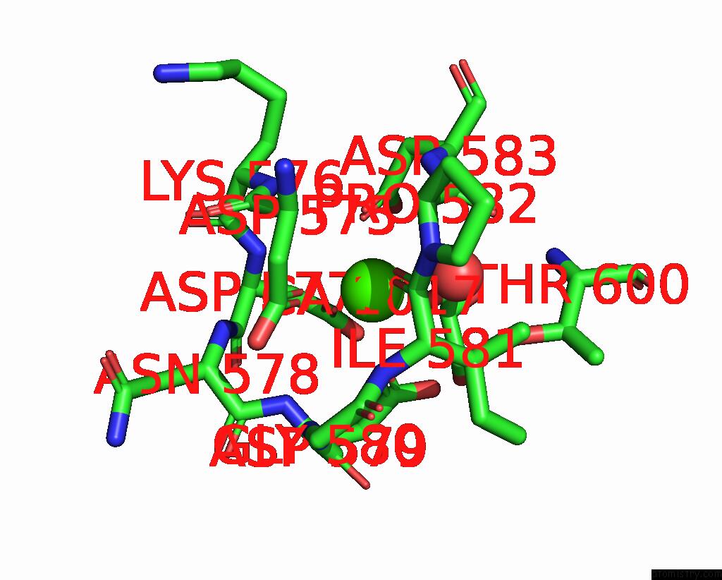
Mono view
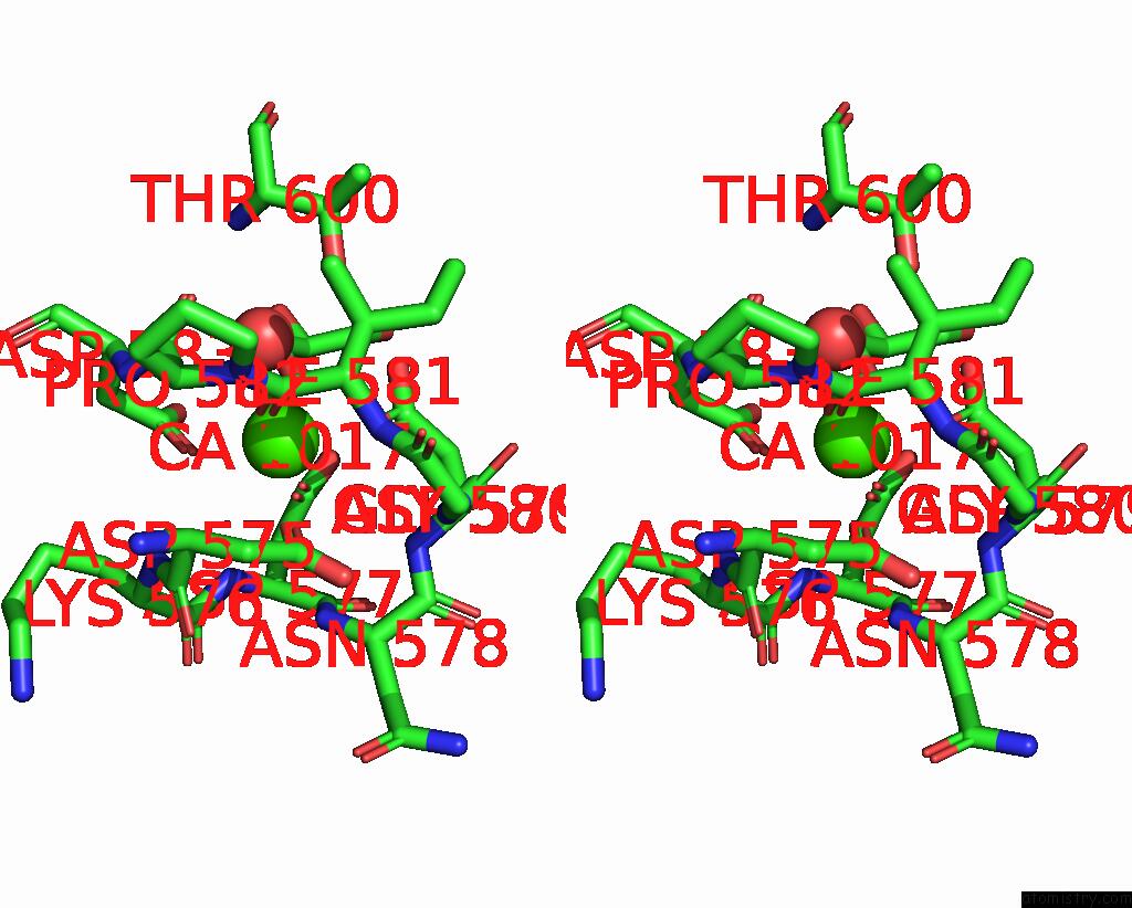
Stereo pair view
Mono view
Stereo pair view
A full contact list of Calcium with other atoms in the Ca binding
site number 1 of Crystal Structure of Thermoanaerobacterium Xylolyticum GH116 Beta- Glucosidase within 5.0Å range:
|
Reference:
R.Charoenwattanasatien,
S.Pengthaisong,
I.Breen,
R.Mutoh,
S.Sansenya,
Y.Hua,
A.Tankrathok,
L.Wu,
C.Songsiriritthigul,
H.Tanaka,
S.J.Williams,
G.J.Davies,
G.Kurisu,
J.R.Ketudat Cairns.
Bacterial Beta-Glucosidase Reveals the Structural and Functional Basis of Genetic Defects in Human Glucocerebrosidase 2 (GBA2). Acs Chem.Biol. V. 11 1891 2016.
ISSN: ESSN 1554-8937
PubMed: 27115290
DOI: 10.1021/ACSCHEMBIO.6B00192
Page generated: Wed Jul 9 04:28:14 2025
ISSN: ESSN 1554-8937
PubMed: 27115290
DOI: 10.1021/ACSCHEMBIO.6B00192
Last articles
Cl in 6E42Cl in 6E72
Cl in 6E6Z
Cl in 6E5Y
Cl in 6E5Z
Cl in 6E5V
Cl in 6E54
Cl in 6E4T
Cl in 6E4U
Cl in 6E4Y