Calcium »
PDB 5cy4-5dau »
5d29 »
Calcium in PDB 5d29: X-Ray Structure of Human Glutamate Carboxypeptidase II (Gcpii) in Complex with A Hydroxamate Inhibitor JHU241
Enzymatic activity of X-Ray Structure of Human Glutamate Carboxypeptidase II (Gcpii) in Complex with A Hydroxamate Inhibitor JHU241
All present enzymatic activity of X-Ray Structure of Human Glutamate Carboxypeptidase II (Gcpii) in Complex with A Hydroxamate Inhibitor JHU241:
3.4.17.21;
3.4.17.21;
Protein crystallography data
The structure of X-Ray Structure of Human Glutamate Carboxypeptidase II (Gcpii) in Complex with A Hydroxamate Inhibitor JHU241, PDB code: 5d29
was solved by
C.Barinka,
Z.Novakova,
J.Pavlicek,
with X-Ray Crystallography technique. A brief refinement statistics is given in the table below:
| Resolution Low / High (Å) | 44.82 / 1.80 |
| Space group | I 2 2 2 |
| Cell size a, b, c (Å), α, β, γ (°) | 100.191, 130.459, 157.196, 90.00, 90.00, 90.00 |
| R / Rfree (%) | 17.4 / 20.5 |
Other elements in 5d29:
The structure of X-Ray Structure of Human Glutamate Carboxypeptidase II (Gcpii) in Complex with A Hydroxamate Inhibitor JHU241 also contains other interesting chemical elements:
| Chlorine | (Cl) | 1 atom |
| Zinc | (Zn) | 2 atoms |
Calcium Binding Sites:
The binding sites of Calcium atom in the X-Ray Structure of Human Glutamate Carboxypeptidase II (Gcpii) in Complex with A Hydroxamate Inhibitor JHU241
(pdb code 5d29). This binding sites where shown within
5.0 Angstroms radius around Calcium atom.
In total only one binding site of Calcium was determined in the X-Ray Structure of Human Glutamate Carboxypeptidase II (Gcpii) in Complex with A Hydroxamate Inhibitor JHU241, PDB code: 5d29:
In total only one binding site of Calcium was determined in the X-Ray Structure of Human Glutamate Carboxypeptidase II (Gcpii) in Complex with A Hydroxamate Inhibitor JHU241, PDB code: 5d29:
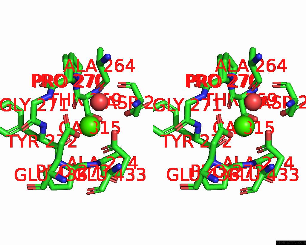
Calcium binding site 1 out of 1 in 5d29
Go back to
Calcium binding site 1 out
of 1 in the X-Ray Structure of Human Glutamate Carboxypeptidase II (Gcpii) in Complex with A Hydroxamate Inhibitor JHU241
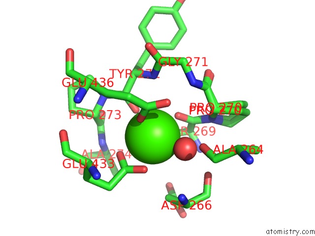
Mono view
Stereo pair view
Mono view
Stereo pair view
A full contact list of Calcium with other atoms in the Ca binding
site number 1 of X-Ray Structure of Human Glutamate Carboxypeptidase II (Gcpii) in Complex with A Hydroxamate Inhibitor JHU241 within 5.0Å range:
|
Reference:
Z.Novakova,
K.Wozniak,
A.Jancarik,
R.Rais,
Y.Wu,
J.Pavlicek,
D.Ferraris,
B.Havlinova,
J.Ptacek,
J.Vavra,
N.Hin,
C.Rojas,
P.Majer,
B.S.Slusher,
T.Tsukamoto,
C.Barinka.
Unprecedented Binding Mode of Hydroxamate-Based Inhibitors of Glutamate Carboxypeptidase II: Structural Characterization and Biological Activity. J.Med.Chem. V. 59 4539 2016.
ISSN: ISSN 0022-2623
PubMed: 27074627
DOI: 10.1021/ACS.JMEDCHEM.5B01806
Page generated: Wed Jul 9 04:58:56 2025
ISSN: ISSN 0022-2623
PubMed: 27074627
DOI: 10.1021/ACS.JMEDCHEM.5B01806
Last articles
K in 1C3OK in 1D7U
K in 1D7S
K in 1D7R
K in 1C38
K in 1CPG
K in 1CPE
K in 1C30
K in 1C7J
K in 1C35