Calcium »
PDB 5cy4-5dau »
5d7f »
Calcium in PDB 5d7f: X-Ray Structure of Ca(2+)-S100B with Human Rage-Derived W72 Peptide
Protein crystallography data
The structure of X-Ray Structure of Ca(2+)-S100B with Human Rage-Derived W72 Peptide, PDB code: 5d7f
was solved by
J.L.Jensen,
C.L.Colbert,
with X-Ray Crystallography technique. A brief refinement statistics is given in the table below:
| Resolution Low / High (Å) | 44.20 / 1.30 |
| Space group | P 1 21 1 |
| Cell size a, b, c (Å), α, β, γ (°) | 35.089, 59.824, 47.563, 90.00, 111.65, 90.00 |
| R / Rfree (%) | 15.7 / 18.3 |
Calcium Binding Sites:
The binding sites of Calcium atom in the X-Ray Structure of Ca(2+)-S100B with Human Rage-Derived W72 Peptide
(pdb code 5d7f). This binding sites where shown within
5.0 Angstroms radius around Calcium atom.
In total 4 binding sites of Calcium where determined in the X-Ray Structure of Ca(2+)-S100B with Human Rage-Derived W72 Peptide, PDB code: 5d7f:
Jump to Calcium binding site number: 1; 2; 3; 4;
In total 4 binding sites of Calcium where determined in the X-Ray Structure of Ca(2+)-S100B with Human Rage-Derived W72 Peptide, PDB code: 5d7f:
Jump to Calcium binding site number: 1; 2; 3; 4;
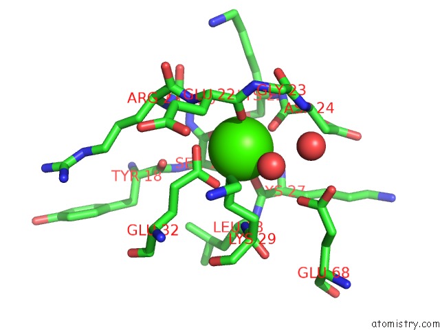
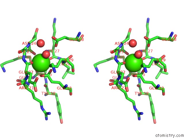
Calcium binding site 1 out of 4 in 5d7f
Go back to
Calcium binding site 1 out
of 4 in the X-Ray Structure of Ca(2+)-S100B with Human Rage-Derived W72 Peptide
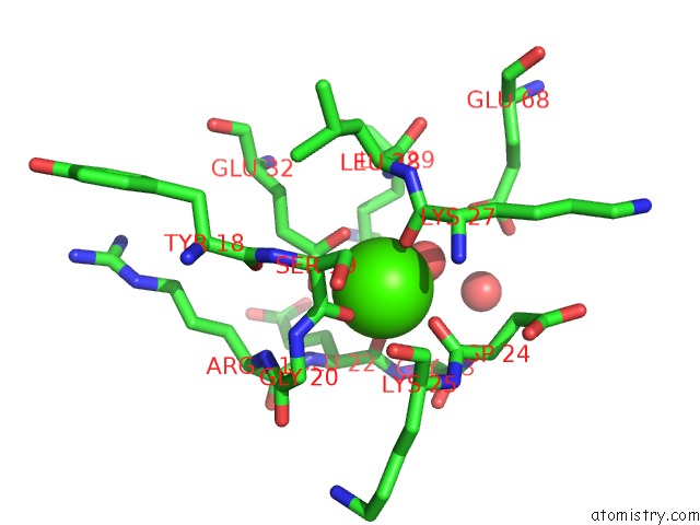
Mono view
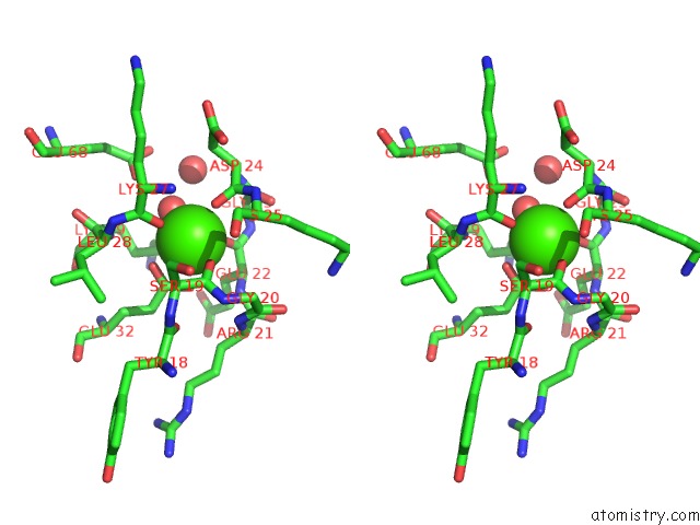
Stereo pair view
Mono view
Stereo pair view
A full contact list of Calcium with other atoms in the Ca binding
site number 1 of X-Ray Structure of Ca(2+)-S100B with Human Rage-Derived W72 Peptide within 5.0Å range:
|
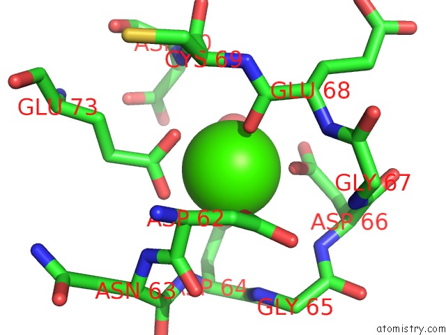
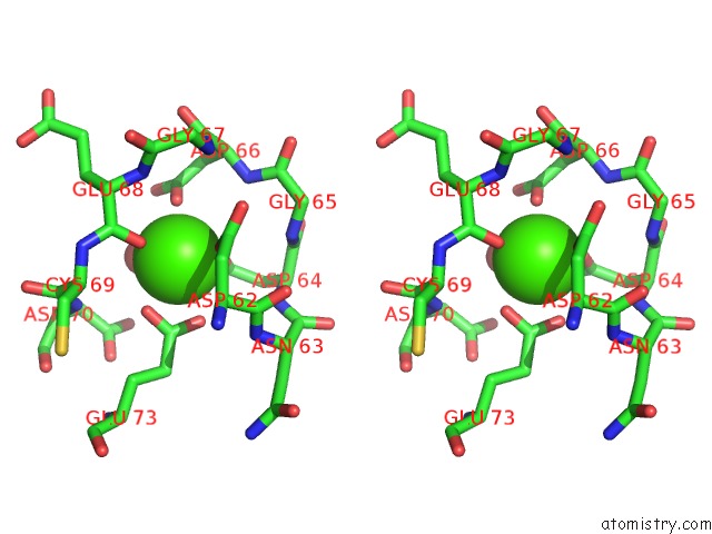
Calcium binding site 2 out of 4 in 5d7f
Go back to
Calcium binding site 2 out
of 4 in the X-Ray Structure of Ca(2+)-S100B with Human Rage-Derived W72 Peptide
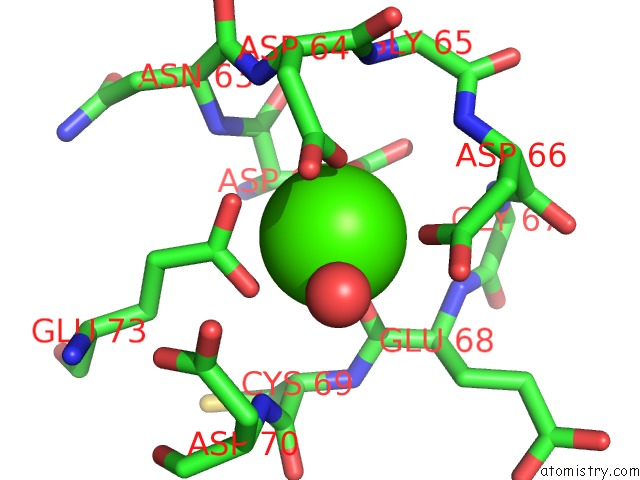
Mono view
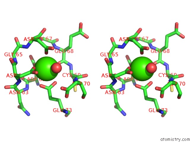
Stereo pair view
Mono view
Stereo pair view
A full contact list of Calcium with other atoms in the Ca binding
site number 2 of X-Ray Structure of Ca(2+)-S100B with Human Rage-Derived W72 Peptide within 5.0Å range:
|
Calcium binding site 3 out of 4 in 5d7f
Go back to
Calcium binding site 3 out
of 4 in the X-Ray Structure of Ca(2+)-S100B with Human Rage-Derived W72 Peptide
Mono view
Stereo pair view
Mono view
Stereo pair view
A full contact list of Calcium with other atoms in the Ca binding
site number 3 of X-Ray Structure of Ca(2+)-S100B with Human Rage-Derived W72 Peptide within 5.0Å range:
|
Calcium binding site 4 out of 4 in 5d7f
Go back to
Calcium binding site 4 out
of 4 in the X-Ray Structure of Ca(2+)-S100B with Human Rage-Derived W72 Peptide
Mono view
Stereo pair view
Mono view
Stereo pair view
A full contact list of Calcium with other atoms in the Ca binding
site number 4 of X-Ray Structure of Ca(2+)-S100B with Human Rage-Derived W72 Peptide within 5.0Å range:
|
Reference:
J.L.Jensen,
C.L.Colbert.
X-Ray Structure of Ca(2+)-S100B with Human Rage-Derived W72 Peptide To Be Published.
Page generated: Wed Jul 9 05:01:51 2025
Last articles
I in 7DCZI in 7D36
I in 7D2V
I in 7D5A
I in 7D2X
I in 7CX9
I in 7CP7
I in 7CRF
I in 7CP6
I in 7BYD