Calcium »
PDB 5cy4-5dau »
5d9o »
Calcium in PDB 5d9o: Crystal Structure of PBGH5A, A Glycoside Hydrolase Family 5 Enzyme From Prevotella Bryantii B14, E280A Mutant in Complex with Cellotetraose
Protein crystallography data
The structure of Crystal Structure of PBGH5A, A Glycoside Hydrolase Family 5 Enzyme From Prevotella Bryantii B14, E280A Mutant in Complex with Cellotetraose, PDB code: 5d9o
was solved by
M.Morar,
P.J.Stogios,
X.Xu,
H.Cui,
R.Di Leo,
V.Yim,
A.Savchenko,
with X-Ray Crystallography technique. A brief refinement statistics is given in the table below:
| Resolution Low / High (Å) | 19.46 / 1.55 |
| Space group | P 1 21 1 |
| Cell size a, b, c (Å), α, β, γ (°) | 49.180, 85.058, 74.978, 90.00, 101.40, 90.00 |
| R / Rfree (%) | 15.3 / 18 |
Calcium Binding Sites:
The binding sites of Calcium atom in the Crystal Structure of PBGH5A, A Glycoside Hydrolase Family 5 Enzyme From Prevotella Bryantii B14, E280A Mutant in Complex with Cellotetraose
(pdb code 5d9o). This binding sites where shown within
5.0 Angstroms radius around Calcium atom.
In total 3 binding sites of Calcium where determined in the Crystal Structure of PBGH5A, A Glycoside Hydrolase Family 5 Enzyme From Prevotella Bryantii B14, E280A Mutant in Complex with Cellotetraose, PDB code: 5d9o:
Jump to Calcium binding site number: 1; 2; 3;
In total 3 binding sites of Calcium where determined in the Crystal Structure of PBGH5A, A Glycoside Hydrolase Family 5 Enzyme From Prevotella Bryantii B14, E280A Mutant in Complex with Cellotetraose, PDB code: 5d9o:
Jump to Calcium binding site number: 1; 2; 3;
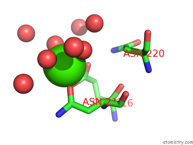
Calcium binding site 1 out of 3 in 5d9o
Go back to
Calcium binding site 1 out
of 3 in the Crystal Structure of PBGH5A, A Glycoside Hydrolase Family 5 Enzyme From Prevotella Bryantii B14, E280A Mutant in Complex with Cellotetraose
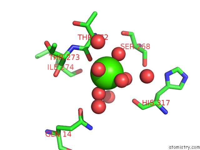
Mono view
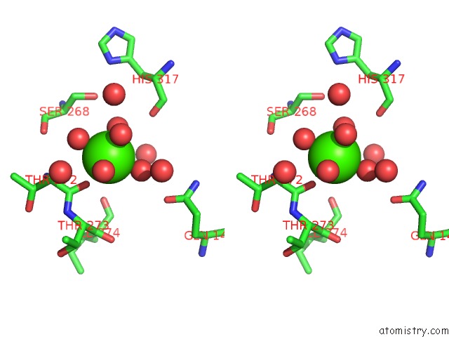
Stereo pair view
Mono view
Stereo pair view
A full contact list of Calcium with other atoms in the Ca binding
site number 1 of Crystal Structure of PBGH5A, A Glycoside Hydrolase Family 5 Enzyme From Prevotella Bryantii B14, E280A Mutant in Complex with Cellotetraose within 5.0Å range:
|
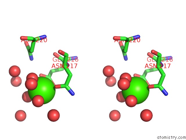
Calcium binding site 2 out of 3 in 5d9o
Go back to
Calcium binding site 2 out
of 3 in the Crystal Structure of PBGH5A, A Glycoside Hydrolase Family 5 Enzyme From Prevotella Bryantii B14, E280A Mutant in Complex with Cellotetraose
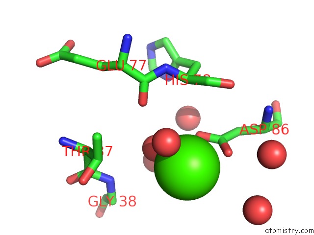
Mono view
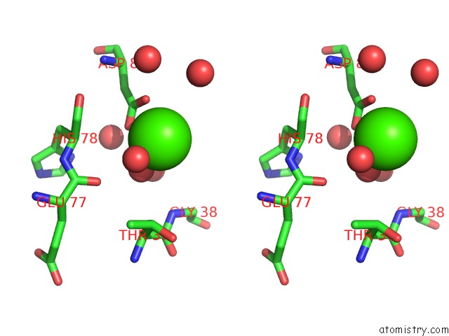
Stereo pair view
Mono view
Stereo pair view
A full contact list of Calcium with other atoms in the Ca binding
site number 2 of Crystal Structure of PBGH5A, A Glycoside Hydrolase Family 5 Enzyme From Prevotella Bryantii B14, E280A Mutant in Complex with Cellotetraose within 5.0Å range:
|
Calcium binding site 3 out of 3 in 5d9o
Go back to
Calcium binding site 3 out
of 3 in the Crystal Structure of PBGH5A, A Glycoside Hydrolase Family 5 Enzyme From Prevotella Bryantii B14, E280A Mutant in Complex with Cellotetraose
Mono view
Stereo pair view
Mono view
Stereo pair view
A full contact list of Calcium with other atoms in the Ca binding
site number 3 of Crystal Structure of PBGH5A, A Glycoside Hydrolase Family 5 Enzyme From Prevotella Bryantii B14, E280A Mutant in Complex with Cellotetraose within 5.0Å range:
|
Reference:
N.Mcgregor,
M.Morar,
T.H.Fenger,
P.Stogios,
N.Lenfant,
V.Yin,
X.Xu,
E.Evdokimova,
H.Cui,
B.Henrissat,
A.Savchenko,
H.Brumer.
Structure-Function Analysis of A Mixed-Linkage Beta-Glucanase/Xyloglucanase From Key Ruminal Bacteroidetes Prevotella Bryantii B14. J.Biol.Chem. 2015.
ISSN: ESSN 1083-351X
PubMed: 26507654
DOI: 10.1074/JBC.M115.691659
Page generated: Wed Jul 9 05:03:42 2025
ISSN: ESSN 1083-351X
PubMed: 26507654
DOI: 10.1074/JBC.M115.691659
Last articles
Mg in 3DTRMg in 3DTA
Mg in 3DSY
Mg in 3DRB
Mg in 3DQW
Mg in 3DR1
Mg in 3DNT
Mg in 3DOF
Mg in 3DOE
Mg in 3DNB