Calcium »
PDB 5dzw-5egt »
5e6u »
Calcium in PDB 5e6u: Structures of Leukocyte Integrin ALB2: the Ai Domain, the Headpiece, and the Pocket For the Internal Ligand
Protein crystallography data
The structure of Structures of Leukocyte Integrin ALB2: the Ai Domain, the Headpiece, and the Pocket For the Internal Ligand, PDB code: 5e6u
was solved by
M.Sen,
T.A.Springer,
with X-Ray Crystallography technique. A brief refinement statistics is given in the table below:
| Resolution Low / High (Å) | 46.44 / 2.50 |
| Space group | P 61 |
| Cell size a, b, c (Å), α, β, γ (°) | 154.980, 154.980, 115.450, 90.00, 90.00, 120.00 |
| R / Rfree (%) | 17.9 / 22.6 |
Other elements in 5e6u:
The structure of Structures of Leukocyte Integrin ALB2: the Ai Domain, the Headpiece, and the Pocket For the Internal Ligand also contains other interesting chemical elements:
| Magnesium | (Mg) | 2 atoms |
| Chlorine | (Cl) | 1 atom |
Calcium Binding Sites:
The binding sites of Calcium atom in the Structures of Leukocyte Integrin ALB2: the Ai Domain, the Headpiece, and the Pocket For the Internal Ligand
(pdb code 5e6u). This binding sites where shown within
5.0 Angstroms radius around Calcium atom.
In total 4 binding sites of Calcium where determined in the Structures of Leukocyte Integrin ALB2: the Ai Domain, the Headpiece, and the Pocket For the Internal Ligand, PDB code: 5e6u:
Jump to Calcium binding site number: 1; 2; 3; 4;
In total 4 binding sites of Calcium where determined in the Structures of Leukocyte Integrin ALB2: the Ai Domain, the Headpiece, and the Pocket For the Internal Ligand, PDB code: 5e6u:
Jump to Calcium binding site number: 1; 2; 3; 4;
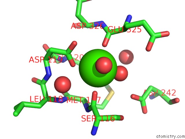
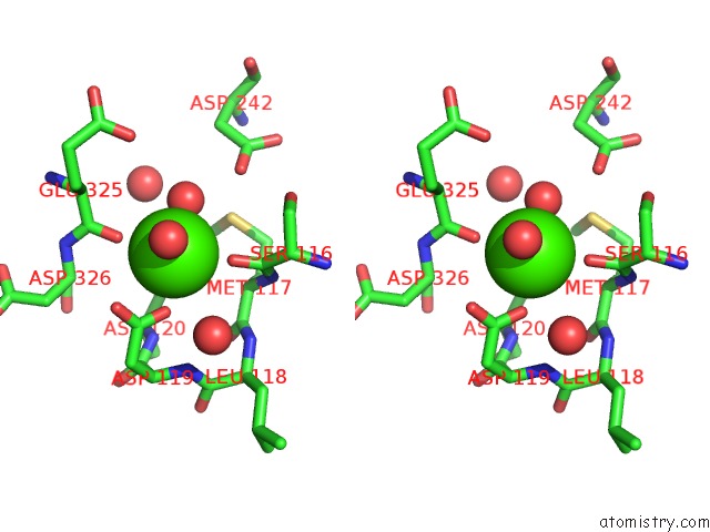
Calcium binding site 1 out of 4 in 5e6u
Go back to
Calcium binding site 1 out
of 4 in the Structures of Leukocyte Integrin ALB2: the Ai Domain, the Headpiece, and the Pocket For the Internal Ligand
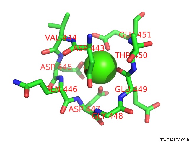
Mono view
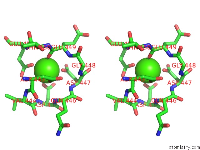
Stereo pair view
Mono view
Stereo pair view
A full contact list of Calcium with other atoms in the Ca binding
site number 1 of Structures of Leukocyte Integrin ALB2: the Ai Domain, the Headpiece, and the Pocket For the Internal Ligand within 5.0Å range:
|
Calcium binding site 2 out of 4 in 5e6u
Go back to
Calcium binding site 2 out
of 4 in the Structures of Leukocyte Integrin ALB2: the Ai Domain, the Headpiece, and the Pocket For the Internal Ligand
Mono view
Stereo pair view
Mono view
Stereo pair view
A full contact list of Calcium with other atoms in the Ca binding
site number 2 of Structures of Leukocyte Integrin ALB2: the Ai Domain, the Headpiece, and the Pocket For the Internal Ligand within 5.0Å range:
|
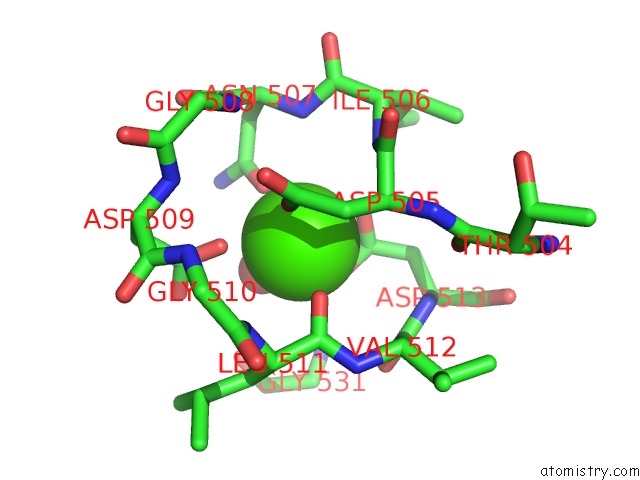
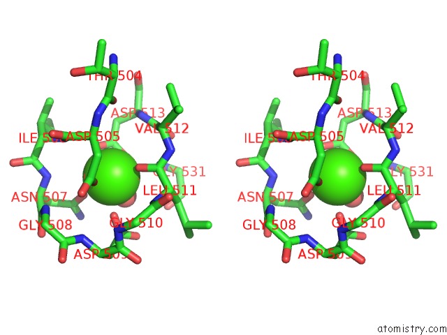
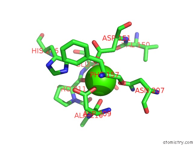
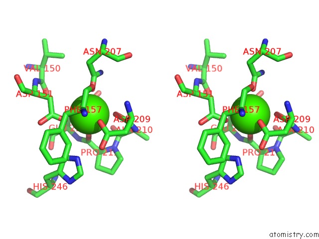
Calcium binding site 3 out of 4 in 5e6u
Go back to
Calcium binding site 3 out
of 4 in the Structures of Leukocyte Integrin ALB2: the Ai Domain, the Headpiece, and the Pocket For the Internal Ligand
Mono view
Stereo pair view
Mono view
Stereo pair view
A full contact list of Calcium with other atoms in the Ca binding
site number 3 of Structures of Leukocyte Integrin ALB2: the Ai Domain, the Headpiece, and the Pocket For the Internal Ligand within 5.0Å range:
|
Calcium binding site 4 out of 4 in 5e6u
Go back to
Calcium binding site 4 out
of 4 in the Structures of Leukocyte Integrin ALB2: the Ai Domain, the Headpiece, and the Pocket For the Internal Ligand
Mono view
Stereo pair view
Mono view
Stereo pair view
A full contact list of Calcium with other atoms in the Ca binding
site number 4 of Structures of Leukocyte Integrin ALB2: the Ai Domain, the Headpiece, and the Pocket For the Internal Ligand within 5.0Å range:
|
Reference:
M.Sen,
T.A.Springer.
Leukocyte Integrin Alpha L Beta 2 Headpiece Structures: the Alpha I Domain, the Pocket For the Internal Ligand, and Concerted Movements of Its Loops. Proc.Natl.Acad.Sci.Usa V. 113 2940 2016.
ISSN: ESSN 1091-6490
PubMed: 26936951
DOI: 10.1073/PNAS.1601379113
Page generated: Wed Jul 9 05:27:07 2025
ISSN: ESSN 1091-6490
PubMed: 26936951
DOI: 10.1073/PNAS.1601379113
Last articles
K in 5SCJK in 5SCI
K in 5SCH
K in 5SCG
K in 5SCF
K in 5SCE
K in 5SCD
K in 5SCA
K in 5SCB
K in 5SC8