Calcium »
PDB 5eym-5fk0 »
5ezf »
Calcium in PDB 5ezf: Racemic Crystal Structures of Pribnow Box Consensus Promoter Sequence (Pbca)
Protein crystallography data
The structure of Racemic Crystal Structures of Pribnow Box Consensus Promoter Sequence (Pbca), PDB code: 5ezf
was solved by
P.K.Mandal,
G.W.Collie,
B.Kauffmann,
S.C.Srivastava,
I.Huc,
with X-Ray Crystallography technique. A brief refinement statistics is given in the table below:
| Resolution Low / High (Å) | 32.86 / 1.65 |
| Space group | P b c a |
| Cell size a, b, c (Å), α, β, γ (°) | 41.736, 39.329, 65.713, 90.00, 90.00, 90.00 |
| R / Rfree (%) | 29.6 / 31.7 |
Calcium Binding Sites:
The binding sites of Calcium atom in the Racemic Crystal Structures of Pribnow Box Consensus Promoter Sequence (Pbca)
(pdb code 5ezf). This binding sites where shown within
5.0 Angstroms radius around Calcium atom.
In total 2 binding sites of Calcium where determined in the Racemic Crystal Structures of Pribnow Box Consensus Promoter Sequence (Pbca), PDB code: 5ezf:
Jump to Calcium binding site number: 1; 2;
In total 2 binding sites of Calcium where determined in the Racemic Crystal Structures of Pribnow Box Consensus Promoter Sequence (Pbca), PDB code: 5ezf:
Jump to Calcium binding site number: 1; 2;
Calcium binding site 1 out of 2 in 5ezf
Go back to
Calcium binding site 1 out
of 2 in the Racemic Crystal Structures of Pribnow Box Consensus Promoter Sequence (Pbca)
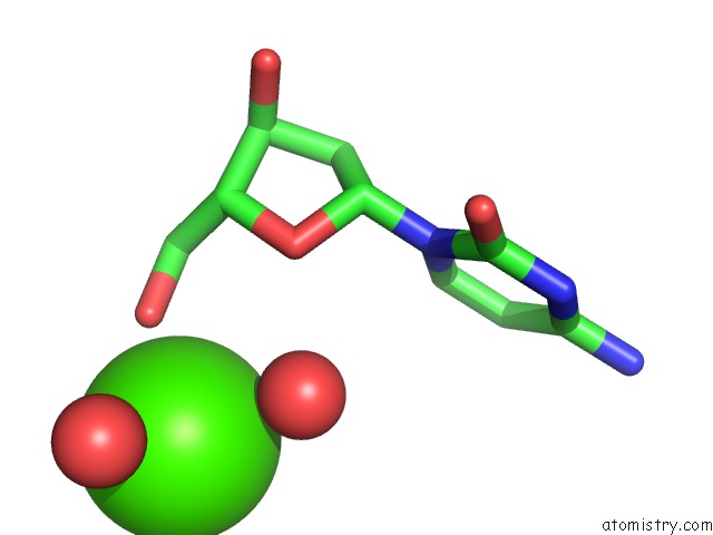
Mono view
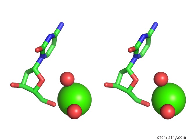
Stereo pair view
Mono view
Stereo pair view
A full contact list of Calcium with other atoms in the Ca binding
site number 1 of Racemic Crystal Structures of Pribnow Box Consensus Promoter Sequence (Pbca) within 5.0Å range:
|
Calcium binding site 2 out of 2 in 5ezf
Go back to
Calcium binding site 2 out
of 2 in the Racemic Crystal Structures of Pribnow Box Consensus Promoter Sequence (Pbca)
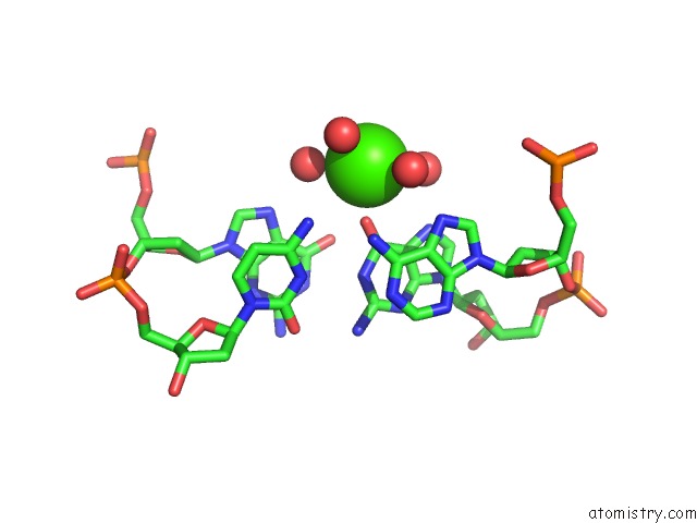
Mono view
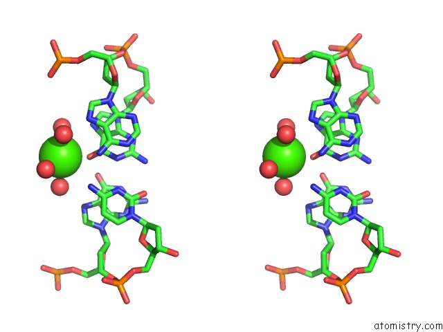
Stereo pair view
Mono view
Stereo pair view
A full contact list of Calcium with other atoms in the Ca binding
site number 2 of Racemic Crystal Structures of Pribnow Box Consensus Promoter Sequence (Pbca) within 5.0Å range:
|
Reference:
P.K.Mandal,
G.W.Collie,
S.C.Srivastava,
B.Kauffmann,
I.Huc.
Structure Elucidation of the Pribnow Box Consensus Promoter Sequence By Racemic Dna Crystallography. Nucleic Acids Res. V. 44 5936 2016.
ISSN: ESSN 1362-4962
PubMed: 27137886
DOI: 10.1093/NAR/GKW367
Page generated: Wed Jul 9 05:47:43 2025
ISSN: ESSN 1362-4962
PubMed: 27137886
DOI: 10.1093/NAR/GKW367
Last articles
Mg in 2XAEMg in 2X9F
Mg in 2X9H
Mg in 2X7D
Mg in 2X7E
Mg in 2X7C
Mg in 2X6S
Mg in 2X6U
Mg in 2X7A
Mg in 2X6V