Calcium »
PDB 5eym-5fk0 »
5f5q »
Calcium in PDB 5f5q: Crystal Structure of Canavalia Virosa Lectin in Complex with Alpha- Methyl-Mannoside
Protein crystallography data
The structure of Crystal Structure of Canavalia Virosa Lectin in Complex with Alpha- Methyl-Mannoside, PDB code: 5f5q
was solved by
V.J.S.Osterne,
J.C.Silva-Filho,
V.R.Pinto-Junior,
M.Q.Santiago,
C.F.Lossio,
P.Delatorre,
K.S.Nascimento,
B.S.Cavada,
with X-Ray Crystallography technique. A brief refinement statistics is given in the table below:
| Resolution Low / High (Å) | 36.99 / 2.52 |
| Space group | P 21 2 21 |
| Cell size a, b, c (Å), α, β, γ (°) | 60.250, 81.740, 86.930, 90.00, 90.00, 90.00 |
| R / Rfree (%) | 18.5 / 25.1 |
Other elements in 5f5q:
The structure of Crystal Structure of Canavalia Virosa Lectin in Complex with Alpha- Methyl-Mannoside also contains other interesting chemical elements:
| Manganese | (Mn) | 2 atoms |
Calcium Binding Sites:
The binding sites of Calcium atom in the Crystal Structure of Canavalia Virosa Lectin in Complex with Alpha- Methyl-Mannoside
(pdb code 5f5q). This binding sites where shown within
5.0 Angstroms radius around Calcium atom.
In total 2 binding sites of Calcium where determined in the Crystal Structure of Canavalia Virosa Lectin in Complex with Alpha- Methyl-Mannoside, PDB code: 5f5q:
Jump to Calcium binding site number: 1; 2;
In total 2 binding sites of Calcium where determined in the Crystal Structure of Canavalia Virosa Lectin in Complex with Alpha- Methyl-Mannoside, PDB code: 5f5q:
Jump to Calcium binding site number: 1; 2;
Calcium binding site 1 out of 2 in 5f5q
Go back to
Calcium binding site 1 out
of 2 in the Crystal Structure of Canavalia Virosa Lectin in Complex with Alpha- Methyl-Mannoside
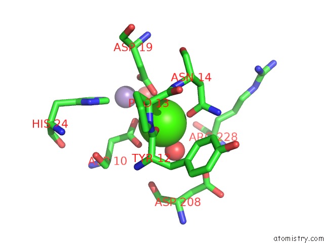
Mono view
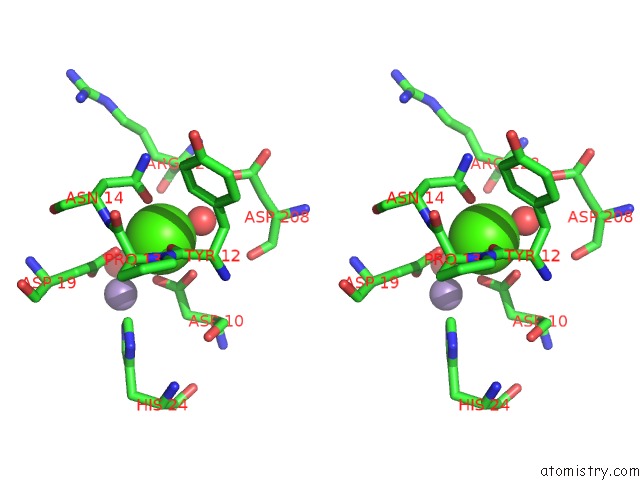
Stereo pair view
Mono view
Stereo pair view
A full contact list of Calcium with other atoms in the Ca binding
site number 1 of Crystal Structure of Canavalia Virosa Lectin in Complex with Alpha- Methyl-Mannoside within 5.0Å range:
|
Calcium binding site 2 out of 2 in 5f5q
Go back to
Calcium binding site 2 out
of 2 in the Crystal Structure of Canavalia Virosa Lectin in Complex with Alpha- Methyl-Mannoside
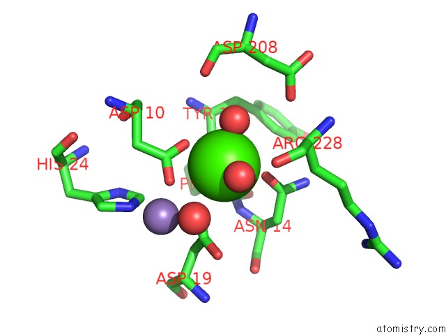
Mono view
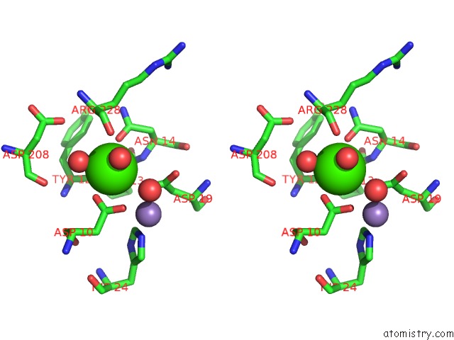
Stereo pair view
Mono view
Stereo pair view
A full contact list of Calcium with other atoms in the Ca binding
site number 2 of Crystal Structure of Canavalia Virosa Lectin in Complex with Alpha- Methyl-Mannoside within 5.0Å range:
|
Reference:
V.J.Osterne,
J.C.Silva-Filho,
M.Q.Santiago,
V.R.Pinto-Junior,
A.C.Almeida,
A.A.Barreto,
I.A.Wolin,
A.P.Nascimento,
R.M.Amorim,
B.A.Rocha,
P.Delatorre,
C.S.Nagano,
R.B.Leal,
A.M.Assreuy,
K.S.Nascimento,
B.S.Cavada.
Structural Characterization of A Lectin From Canavalia Virosa Seeds with Inflammatory and Cytotoxic Activities. Int.J.Biol.Macromol. V. 94 271 2016.
ISSN: ISSN 0141-8130
PubMed: 27737777
DOI: 10.1016/J.IJBIOMAC.2016.10.020
Page generated: Wed Jul 9 05:52:03 2025
ISSN: ISSN 0141-8130
PubMed: 27737777
DOI: 10.1016/J.IJBIOMAC.2016.10.020
Last articles
Mg in 2BJ3Mg in 2BIF
Mg in 2BIA
Mg in 2BI5
Mg in 2BI9
Mg in 2BI3
Mg in 2BI2
Mg in 2BI1
Mg in 2BHZ
Mg in 2BHX