Calcium »
PDB 5fl0-5g39 »
5ful »
Calcium in PDB 5ful: Crystal Structure of Mus Musculus Protein Arginine Methyltransferase 2 with Sah
Enzymatic activity of Crystal Structure of Mus Musculus Protein Arginine Methyltransferase 2 with Sah
All present enzymatic activity of Crystal Structure of Mus Musculus Protein Arginine Methyltransferase 2 with Sah:
2.1.1.125;
2.1.1.125;
Protein crystallography data
The structure of Crystal Structure of Mus Musculus Protein Arginine Methyltransferase 2 with Sah, PDB code: 5ful
was solved by
V.Cura,
N.Troffer-Charlier,
N.Marechal,
L.Bonnefond,
J.Cavarelli,
with X-Ray Crystallography technique. A brief refinement statistics is given in the table below:
| Resolution Low / High (Å) | 43.26 / 1.89 |
| Space group | C 2 2 21 |
| Cell size a, b, c (Å), α, β, γ (°) | 65.507, 115.414, 132.963, 90.00, 90.00, 90.00 |
| R / Rfree (%) | 16.6 / 19.2 |
Other elements in 5ful:
The structure of Crystal Structure of Mus Musculus Protein Arginine Methyltransferase 2 with Sah also contains other interesting chemical elements:
| Chlorine | (Cl) | 1 atom |
Calcium Binding Sites:
The binding sites of Calcium atom in the Crystal Structure of Mus Musculus Protein Arginine Methyltransferase 2 with Sah
(pdb code 5ful). This binding sites where shown within
5.0 Angstroms radius around Calcium atom.
In total 2 binding sites of Calcium where determined in the Crystal Structure of Mus Musculus Protein Arginine Methyltransferase 2 with Sah, PDB code: 5ful:
Jump to Calcium binding site number: 1; 2;
In total 2 binding sites of Calcium where determined in the Crystal Structure of Mus Musculus Protein Arginine Methyltransferase 2 with Sah, PDB code: 5ful:
Jump to Calcium binding site number: 1; 2;
Calcium binding site 1 out of 2 in 5ful
Go back to
Calcium binding site 1 out
of 2 in the Crystal Structure of Mus Musculus Protein Arginine Methyltransferase 2 with Sah

Mono view
Stereo pair view
Mono view
Stereo pair view
A full contact list of Calcium with other atoms in the Ca binding
site number 1 of Crystal Structure of Mus Musculus Protein Arginine Methyltransferase 2 with Sah within 5.0Å range:
|
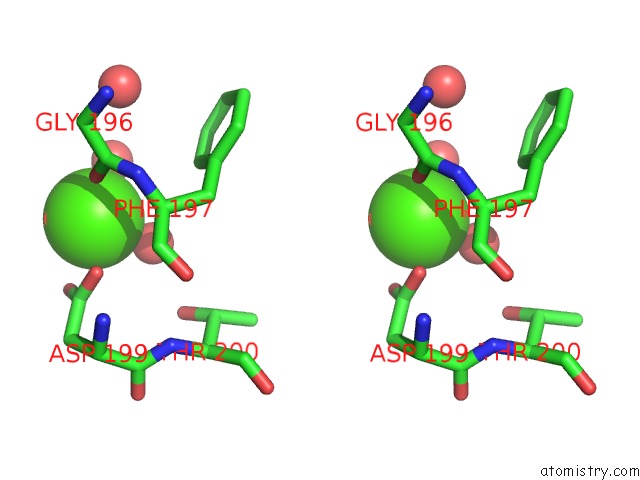
Calcium binding site 2 out of 2 in 5ful
Go back to
Calcium binding site 2 out
of 2 in the Crystal Structure of Mus Musculus Protein Arginine Methyltransferase 2 with Sah

Mono view

Stereo pair view
Mono view
Stereo pair view
A full contact list of Calcium with other atoms in the Ca binding
site number 2 of Crystal Structure of Mus Musculus Protein Arginine Methyltransferase 2 with Sah within 5.0Å range:
|
Reference:
V.Cura,
N.Marechal,
N.Troffer-Charlier,
J.M.Strub,
M.J.Van Haren,
N.I.Martin,
S.Cianferani,
L.Bonnefond,
J.Cavarelli.
Structural Studies of Protein Arginine Methyltransferase 2 Reveal Its Interactions with Potential Substrates and Inhibitors. Febs J. V. 284 77 2017.
ISSN: ISSN 1742-4658
PubMed: 27879050
DOI: 10.1111/FEBS.13953
Page generated: Wed Jul 9 06:02:35 2025
ISSN: ISSN 1742-4658
PubMed: 27879050
DOI: 10.1111/FEBS.13953
Last articles
K in 4EVYK in 4EOU
K in 4ETM
K in 4ESK
K in 4ES8
K in 4ERT
K in 4ERD
K in 4ENC
K in 4EK1
K in 4ENB