Calcium »
PDB 5hcb-5hsa »
5hkj »
Calcium in PDB 5hkj: Single Chain Recombinant Globular Head of the Complement System Protein C1Q
Protein crystallography data
The structure of Single Chain Recombinant Globular Head of the Complement System Protein C1Q, PDB code: 5hkj
was solved by
C.P.Moreau,
C.Gaboriaud,
with X-Ray Crystallography technique. A brief refinement statistics is given in the table below:
| Resolution Low / High (Å) | 42.82 / 1.35 |
| Space group | C 1 2 1 |
| Cell size a, b, c (Å), α, β, γ (°) | 81.115, 52.729, 89.930, 90.00, 115.22, 90.00 |
| R / Rfree (%) | 17.5 / 20 |
Calcium Binding Sites:
The binding sites of Calcium atom in the Single Chain Recombinant Globular Head of the Complement System Protein C1Q
(pdb code 5hkj). This binding sites where shown within
5.0 Angstroms radius around Calcium atom.
In total only one binding site of Calcium was determined in the Single Chain Recombinant Globular Head of the Complement System Protein C1Q, PDB code: 5hkj:
In total only one binding site of Calcium was determined in the Single Chain Recombinant Globular Head of the Complement System Protein C1Q, PDB code: 5hkj:
Calcium binding site 1 out of 1 in 5hkj
Go back to
Calcium binding site 1 out
of 1 in the Single Chain Recombinant Globular Head of the Complement System Protein C1Q
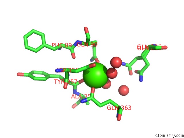
Mono view
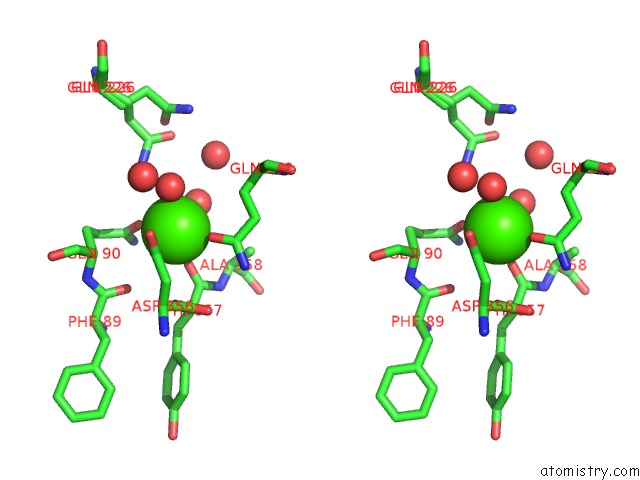
Stereo pair view
Mono view
Stereo pair view
A full contact list of Calcium with other atoms in the Ca binding
site number 1 of Single Chain Recombinant Globular Head of the Complement System Protein C1Q within 5.0Å range:
|
Reference:
C.Moreau,
I.Bally,
A.Chouquet,
B.Bottazzi,
B.Ghebrehiwet,
C.Gaboriaud,
N.Thielens.
Structural and Functional Characterization of A Single-Chain Form of the Recognition Domain of Complement Protein C1Q. Front Immunol V. 7 79 2016.
ISSN: ESSN 1664-3224
PubMed: 26973654
DOI: 10.3389/FIMMU.2016.00079
Page generated: Wed Jul 9 06:29:14 2025
ISSN: ESSN 1664-3224
PubMed: 26973654
DOI: 10.3389/FIMMU.2016.00079
Last articles
Mg in 5XTMMg in 5XUT
Mg in 5XUS
Mg in 5XUJ
Mg in 5XUI
Mg in 5XU1
Mg in 5XT8
Mg in 5XT2
Mg in 5XR7
Mg in 5XR6