Calcium »
PDB 5hcb-5hsa »
5hm4 »
Calcium in PDB 5hm4: Crystal Structure of Oligopeptide Abc Transporter, Periplasmic Oligopeptide-Binding Protein (TM1226) From Thermotoga Maritima at 2.0 A Resolution
Protein crystallography data
The structure of Crystal Structure of Oligopeptide Abc Transporter, Periplasmic Oligopeptide-Binding Protein (TM1226) From Thermotoga Maritima at 2.0 A Resolution, PDB code: 5hm4
was solved by
X.Lu,
S.Ghimire-Rijal,
D.A.A.Myles,
M.J.Cuneo,
with X-Ray Crystallography technique. A brief refinement statistics is given in the table below:
| Resolution Low / High (Å) | 40.44 / 2.00 |
| Space group | P 21 2 21 |
| Cell size a, b, c (Å), α, β, γ (°) | 51.256, 65.795, 185.658, 90.00, 90.00, 90.00 |
| R / Rfree (%) | 17.3 / 20.9 |
Calcium Binding Sites:
The binding sites of Calcium atom in the Crystal Structure of Oligopeptide Abc Transporter, Periplasmic Oligopeptide-Binding Protein (TM1226) From Thermotoga Maritima at 2.0 A Resolution
(pdb code 5hm4). This binding sites where shown within
5.0 Angstroms radius around Calcium atom.
In total only one binding site of Calcium was determined in the Crystal Structure of Oligopeptide Abc Transporter, Periplasmic Oligopeptide-Binding Protein (TM1226) From Thermotoga Maritima at 2.0 A Resolution, PDB code: 5hm4:
In total only one binding site of Calcium was determined in the Crystal Structure of Oligopeptide Abc Transporter, Periplasmic Oligopeptide-Binding Protein (TM1226) From Thermotoga Maritima at 2.0 A Resolution, PDB code: 5hm4:
Calcium binding site 1 out of 1 in 5hm4
Go back to
Calcium binding site 1 out
of 1 in the Crystal Structure of Oligopeptide Abc Transporter, Periplasmic Oligopeptide-Binding Protein (TM1226) From Thermotoga Maritima at 2.0 A Resolution
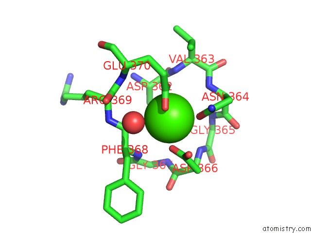
Mono view
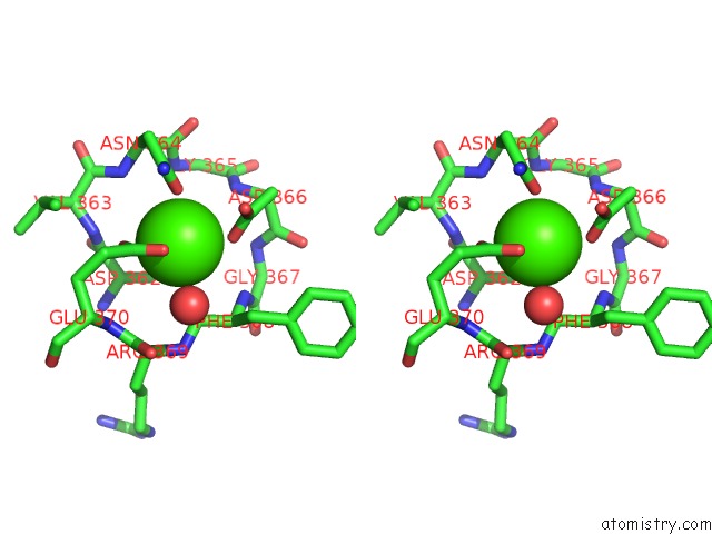
Stereo pair view
Mono view
Stereo pair view
A full contact list of Calcium with other atoms in the Ca binding
site number 1 of Crystal Structure of Oligopeptide Abc Transporter, Periplasmic Oligopeptide-Binding Protein (TM1226) From Thermotoga Maritima at 2.0 A Resolution within 5.0Å range:
|
Reference:
L.Li,
S.Ghimire-Rijal,
S.L.Lucas,
C.B.Stanley,
E.Wright,
P.K.Agarwal,
D.A.Myles,
M.J.Cuneo.
Periplasmic Binding Protein Dimer Has A Second Allosteric Event Tied to Ligand Binding. Biochemistry V. 56 5328 2017.
ISSN: ISSN 1520-4995
PubMed: 28876049
DOI: 10.1021/ACS.BIOCHEM.7B00657
Page generated: Wed Jul 9 06:30:30 2025
ISSN: ISSN 1520-4995
PubMed: 28876049
DOI: 10.1021/ACS.BIOCHEM.7B00657
Last articles
Mg in 6EE4Mg in 6EEC
Mg in 6EE8
Mg in 6EE6
Mg in 6EDW
Mg in 6EE3
Mg in 6EE1
Mg in 6EAC
Mg in 6ED2
Mg in 6EDT