Calcium »
PDB 5jap-5jrc »
5jmo »
Calcium in PDB 5jmo: X-Ray Structure of Furin in Complex with the Inhibitory Antibody NB14
Enzymatic activity of X-Ray Structure of Furin in Complex with the Inhibitory Antibody NB14
All present enzymatic activity of X-Ray Structure of Furin in Complex with the Inhibitory Antibody NB14:
3.4.21.75;
3.4.21.75;
Protein crystallography data
The structure of X-Ray Structure of Furin in Complex with the Inhibitory Antibody NB14, PDB code: 5jmo
was solved by
S.O.Dahms,
M.E.Than,
with X-Ray Crystallography technique. A brief refinement statistics is given in the table below:
| Resolution Low / High (Å) | 66.38 / 2.00 |
| Space group | P 21 2 21 |
| Cell size a, b, c (Å), α, β, γ (°) | 169.775, 50.039, 144.244, 90.00, 90.00, 90.00 |
| R / Rfree (%) | 16.3 / 19.7 |
Other elements in 5jmo:
The structure of X-Ray Structure of Furin in Complex with the Inhibitory Antibody NB14 also contains other interesting chemical elements:
| Sodium | (Na) | 2 atoms |
Calcium Binding Sites:
The binding sites of Calcium atom in the X-Ray Structure of Furin in Complex with the Inhibitory Antibody NB14
(pdb code 5jmo). This binding sites where shown within
5.0 Angstroms radius around Calcium atom.
In total 6 binding sites of Calcium where determined in the X-Ray Structure of Furin in Complex with the Inhibitory Antibody NB14, PDB code: 5jmo:
Jump to Calcium binding site number: 1; 2; 3; 4; 5; 6;
In total 6 binding sites of Calcium where determined in the X-Ray Structure of Furin in Complex with the Inhibitory Antibody NB14, PDB code: 5jmo:
Jump to Calcium binding site number: 1; 2; 3; 4; 5; 6;
Calcium binding site 1 out of 6 in 5jmo
Go back to
Calcium binding site 1 out
of 6 in the X-Ray Structure of Furin in Complex with the Inhibitory Antibody NB14

Mono view
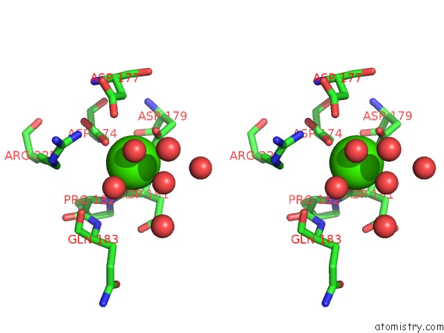
Stereo pair view
Mono view
Stereo pair view
A full contact list of Calcium with other atoms in the Ca binding
site number 1 of X-Ray Structure of Furin in Complex with the Inhibitory Antibody NB14 within 5.0Å range:
|
Calcium binding site 2 out of 6 in 5jmo
Go back to
Calcium binding site 2 out
of 6 in the X-Ray Structure of Furin in Complex with the Inhibitory Antibody NB14

Mono view

Stereo pair view
Mono view
Stereo pair view
A full contact list of Calcium with other atoms in the Ca binding
site number 2 of X-Ray Structure of Furin in Complex with the Inhibitory Antibody NB14 within 5.0Å range:
|
Calcium binding site 3 out of 6 in 5jmo
Go back to
Calcium binding site 3 out
of 6 in the X-Ray Structure of Furin in Complex with the Inhibitory Antibody NB14

Mono view

Stereo pair view
Mono view
Stereo pair view
A full contact list of Calcium with other atoms in the Ca binding
site number 3 of X-Ray Structure of Furin in Complex with the Inhibitory Antibody NB14 within 5.0Å range:
|
Calcium binding site 4 out of 6 in 5jmo
Go back to
Calcium binding site 4 out
of 6 in the X-Ray Structure of Furin in Complex with the Inhibitory Antibody NB14

Mono view

Stereo pair view
Mono view
Stereo pair view
A full contact list of Calcium with other atoms in the Ca binding
site number 4 of X-Ray Structure of Furin in Complex with the Inhibitory Antibody NB14 within 5.0Å range:
|
Calcium binding site 5 out of 6 in 5jmo
Go back to
Calcium binding site 5 out
of 6 in the X-Ray Structure of Furin in Complex with the Inhibitory Antibody NB14

Mono view

Stereo pair view
Mono view
Stereo pair view
A full contact list of Calcium with other atoms in the Ca binding
site number 5 of X-Ray Structure of Furin in Complex with the Inhibitory Antibody NB14 within 5.0Å range:
|
Calcium binding site 6 out of 6 in 5jmo
Go back to
Calcium binding site 6 out
of 6 in the X-Ray Structure of Furin in Complex with the Inhibitory Antibody NB14

Mono view

Stereo pair view
Mono view
Stereo pair view
A full contact list of Calcium with other atoms in the Ca binding
site number 6 of X-Ray Structure of Furin in Complex with the Inhibitory Antibody NB14 within 5.0Å range:
|
Reference:
S.O.Dahms,
J.W.Creemers,
Y.Schaub,
G.P.Bourenkov,
T.Zogg,
H.Brandstetter,
M.E.Than.
The Structure of A Furin-Antibody Complex Explains Non-Competitive Inhibition By Steric Exclusion of Substrate Conformers. Sci Rep V. 6 34303 2016.
ISSN: ESSN 2045-2322
PubMed: 27670069
DOI: 10.1038/SREP34303
Page generated: Wed Jul 9 07:08:13 2025
ISSN: ESSN 2045-2322
PubMed: 27670069
DOI: 10.1038/SREP34303
Last articles
K in 4X3ZK in 4X3M
K in 4X16
K in 4X17
K in 4X3K
K in 4X12
K in 4X14
K in 4X15
K in 4X13
K in 4X11