Calcium »
PDB 5jrj-5kaf »
5k5s »
Calcium in PDB 5k5s: Crystal Structure of the Active Form of Human Calcium-Sensing Receptor Extracellular Domain
Protein crystallography data
The structure of Crystal Structure of the Active Form of Human Calcium-Sensing Receptor Extracellular Domain, PDB code: 5k5s
was solved by
Y.Geng,
L.Mosyak,
I.Kurinov,
H.Zuo,
E.Sturchler,
T.C.Cheng,
P.Subramanyam,
A.P.Brown,
S.C.Brennan,
H.-C.Mun,
M.Bush,
Y.Chen,
T.Nguyen,
B.Cao,
D.Chang,
M.Quick,
A.Conigrave,
H.M.Colecraft,
P.Mcdonald,
Q.R.Fan,
with X-Ray Crystallography technique. A brief refinement statistics is given in the table below:
| Resolution Low / High (Å) | 38.07 / 2.60 |
| Space group | C 1 2 1 |
| Cell size a, b, c (Å), α, β, γ (°) | 107.660, 127.450, 146.770, 90.00, 108.72, 90.00 |
| R / Rfree (%) | 21.1 / 22.2 |
Calcium Binding Sites:
The binding sites of Calcium atom in the Crystal Structure of the Active Form of Human Calcium-Sensing Receptor Extracellular Domain
(pdb code 5k5s). This binding sites where shown within
5.0 Angstroms radius around Calcium atom.
In total 8 binding sites of Calcium where determined in the Crystal Structure of the Active Form of Human Calcium-Sensing Receptor Extracellular Domain, PDB code: 5k5s:
Jump to Calcium binding site number: 1; 2; 3; 4; 5; 6; 7; 8;
In total 8 binding sites of Calcium where determined in the Crystal Structure of the Active Form of Human Calcium-Sensing Receptor Extracellular Domain, PDB code: 5k5s:
Jump to Calcium binding site number: 1; 2; 3; 4; 5; 6; 7; 8;
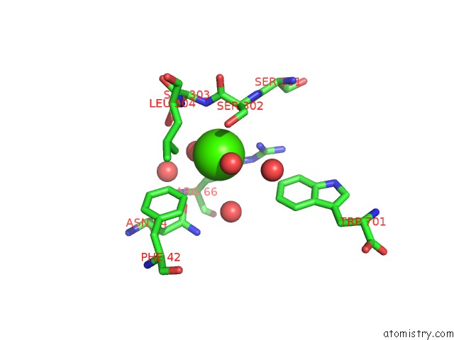
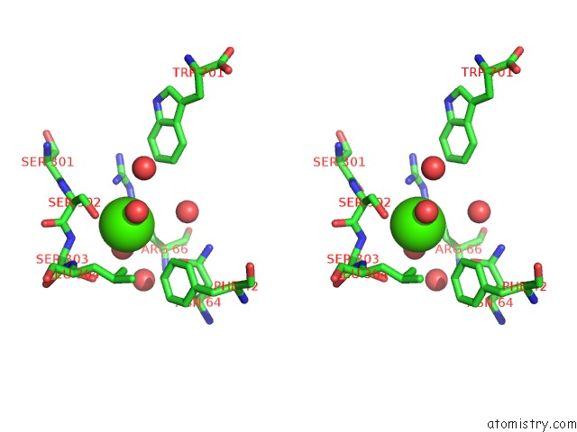
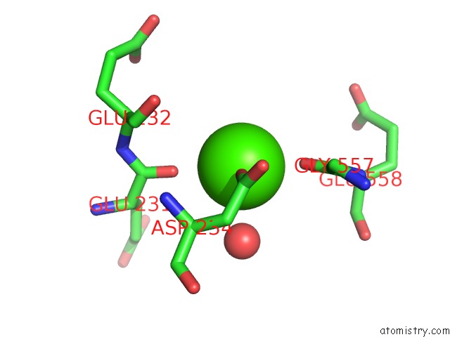
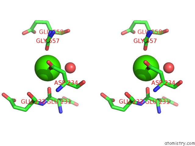
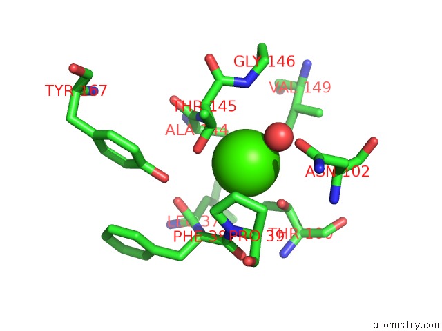
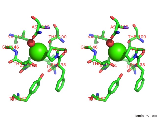
Calcium binding site 1 out of 8 in 5k5s
Go back to
Calcium binding site 1 out
of 8 in the Crystal Structure of the Active Form of Human Calcium-Sensing Receptor Extracellular Domain
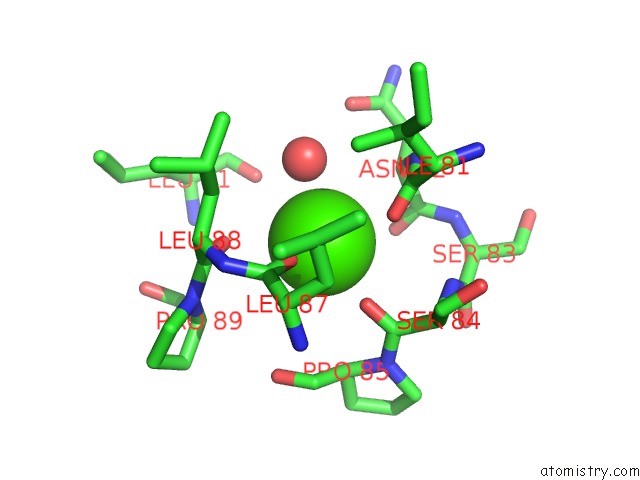
Mono view
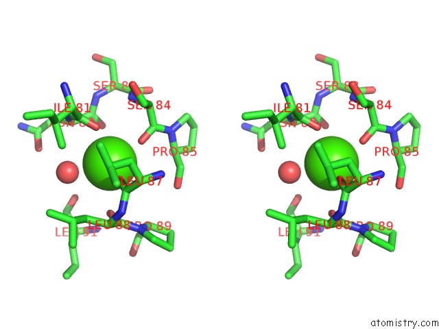
Stereo pair view
Mono view
Stereo pair view
A full contact list of Calcium with other atoms in the Ca binding
site number 1 of Crystal Structure of the Active Form of Human Calcium-Sensing Receptor Extracellular Domain within 5.0Å range:
|
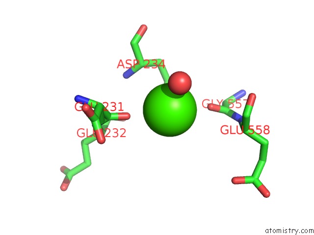
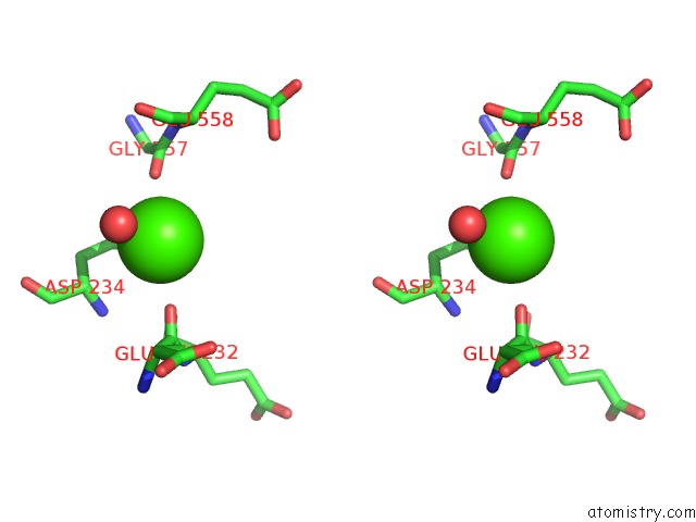
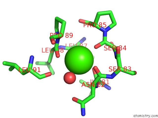
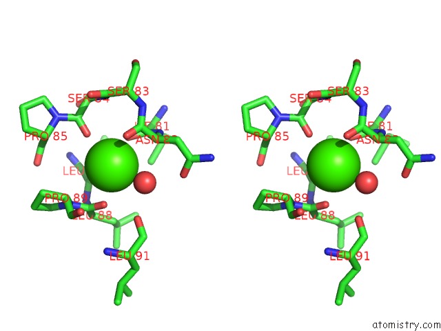
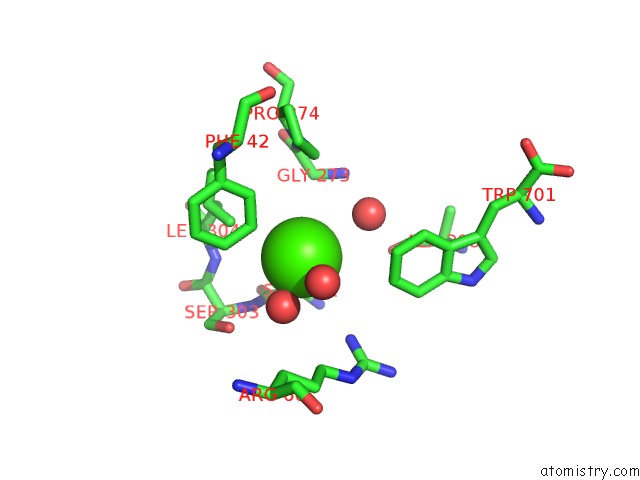
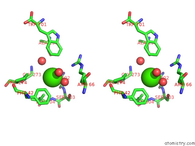
Calcium binding site 2 out of 8 in 5k5s
Go back to
Calcium binding site 2 out
of 8 in the Crystal Structure of the Active Form of Human Calcium-Sensing Receptor Extracellular Domain
Mono view
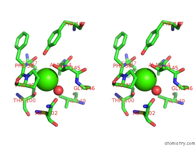
Stereo pair view
Mono view
Stereo pair view
A full contact list of Calcium with other atoms in the Ca binding
site number 2 of Crystal Structure of the Active Form of Human Calcium-Sensing Receptor Extracellular Domain within 5.0Å range:
|
Calcium binding site 3 out of 8 in 5k5s
Go back to
Calcium binding site 3 out
of 8 in the Crystal Structure of the Active Form of Human Calcium-Sensing Receptor Extracellular Domain
Mono view
Stereo pair view
Mono view
Stereo pair view
A full contact list of Calcium with other atoms in the Ca binding
site number 3 of Crystal Structure of the Active Form of Human Calcium-Sensing Receptor Extracellular Domain within 5.0Å range:
|
Calcium binding site 4 out of 8 in 5k5s
Go back to
Calcium binding site 4 out
of 8 in the Crystal Structure of the Active Form of Human Calcium-Sensing Receptor Extracellular Domain
Mono view
Stereo pair view
Mono view
Stereo pair view
A full contact list of Calcium with other atoms in the Ca binding
site number 4 of Crystal Structure of the Active Form of Human Calcium-Sensing Receptor Extracellular Domain within 5.0Å range:
|
Calcium binding site 5 out of 8 in 5k5s
Go back to
Calcium binding site 5 out
of 8 in the Crystal Structure of the Active Form of Human Calcium-Sensing Receptor Extracellular Domain
Mono view
Stereo pair view
Mono view
Stereo pair view
A full contact list of Calcium with other atoms in the Ca binding
site number 5 of Crystal Structure of the Active Form of Human Calcium-Sensing Receptor Extracellular Domain within 5.0Å range:
|
Calcium binding site 6 out of 8 in 5k5s
Go back to
Calcium binding site 6 out
of 8 in the Crystal Structure of the Active Form of Human Calcium-Sensing Receptor Extracellular Domain
Mono view
Stereo pair view
Mono view
Stereo pair view
A full contact list of Calcium with other atoms in the Ca binding
site number 6 of Crystal Structure of the Active Form of Human Calcium-Sensing Receptor Extracellular Domain within 5.0Å range:
|
Calcium binding site 7 out of 8 in 5k5s
Go back to
Calcium binding site 7 out
of 8 in the Crystal Structure of the Active Form of Human Calcium-Sensing Receptor Extracellular Domain
Mono view
Stereo pair view
Mono view
Stereo pair view
A full contact list of Calcium with other atoms in the Ca binding
site number 7 of Crystal Structure of the Active Form of Human Calcium-Sensing Receptor Extracellular Domain within 5.0Å range:
|
Calcium binding site 8 out of 8 in 5k5s
Go back to
Calcium binding site 8 out
of 8 in the Crystal Structure of the Active Form of Human Calcium-Sensing Receptor Extracellular Domain
Mono view
Stereo pair view
Mono view
Stereo pair view
A full contact list of Calcium with other atoms in the Ca binding
site number 8 of Crystal Structure of the Active Form of Human Calcium-Sensing Receptor Extracellular Domain within 5.0Å range:
|
Reference:
Y.Geng,
L.Mosyak,
I.Kurinov,
H.Zuo,
E.Sturchler,
T.C.Cheng,
P.Subramanyam,
A.P.Brown,
S.C.Brennan,
H.C.Mun,
M.Bush,
Y.Chen,
T.X.Nguyen,
B.Cao,
D.D.Chang,
M.Quick,
A.D.Conigrave,
H.M.Colecraft,
P.Mcdonald,
Q.R.Fan.
Structural Mechanism of Ligand Activation in Human Calcium-Sensing Receptor. Elife V. 5 2016.
ISSN: ESSN 2050-084X
PubMed: 27434672
DOI: 10.7554/ELIFE.13662
Page generated: Wed Jul 9 07:17:54 2025
ISSN: ESSN 2050-084X
PubMed: 27434672
DOI: 10.7554/ELIFE.13662
Last articles
Mn in 9LJUMn in 9LJW
Mn in 9LJS
Mn in 9LJR
Mn in 9LJT
Mn in 9LJV
Mg in 9UA2
Mg in 9R96
Mg in 9VM1
Mg in 9P01