Calcium »
PDB 5jrj-5kaf »
5k7s »
Calcium in PDB 5k7s: Microed Structure of Proteinase K at 1.6 A Resolution
Enzymatic activity of Microed Structure of Proteinase K at 1.6 A Resolution
All present enzymatic activity of Microed Structure of Proteinase K at 1.6 A Resolution:
3.4.21.64;
3.4.21.64;
Calcium Binding Sites:
The binding sites of Calcium atom in the Microed Structure of Proteinase K at 1.6 A Resolution
(pdb code 5k7s). This binding sites where shown within
5.0 Angstroms radius around Calcium atom.
In total 2 binding sites of Calcium where determined in the Microed Structure of Proteinase K at 1.6 A Resolution, PDB code: 5k7s:
Jump to Calcium binding site number: 1; 2;
In total 2 binding sites of Calcium where determined in the Microed Structure of Proteinase K at 1.6 A Resolution, PDB code: 5k7s:
Jump to Calcium binding site number: 1; 2;
Calcium binding site 1 out of 2 in 5k7s
Go back to
Calcium binding site 1 out
of 2 in the Microed Structure of Proteinase K at 1.6 A Resolution
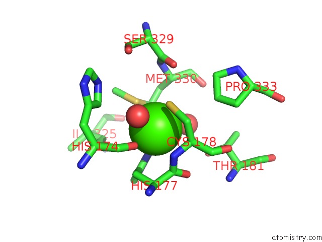
Mono view
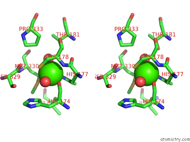
Stereo pair view
Mono view
Stereo pair view
A full contact list of Calcium with other atoms in the Ca binding
site number 1 of Microed Structure of Proteinase K at 1.6 A Resolution within 5.0Å range:
|
Calcium binding site 2 out of 2 in 5k7s
Go back to
Calcium binding site 2 out
of 2 in the Microed Structure of Proteinase K at 1.6 A Resolution
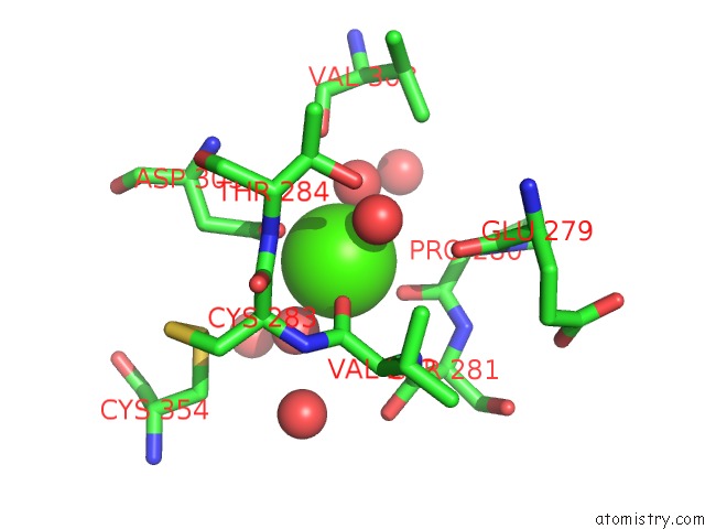
Mono view
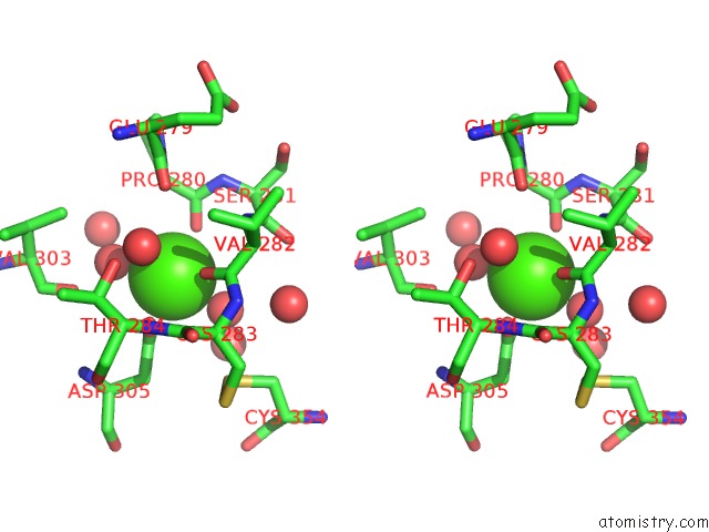
Stereo pair view
Mono view
Stereo pair view
A full contact list of Calcium with other atoms in the Ca binding
site number 2 of Microed Structure of Proteinase K at 1.6 A Resolution within 5.0Å range:
|
Reference:
M.J.De La Cruz,
J.Hattne,
D.Shi,
P.Seidler,
J.Rodriguez,
F.E.Reyes,
M.R.Sawaya,
D.Cascio,
S.C.Weiss,
S.K.Kim,
C.S.Hinck,
A.P.Hinck,
G.Calero,
D.Eisenberg,
T.Gonen.
Atomic-Resolution Structures From Fragmented Protein Crystals with the Cryoem Method Microed. Nat. Methods V. 14 399 2017.
ISSN: ESSN 1548-7105
PubMed: 28192420
DOI: 10.1038/NMETH.4178
Page generated: Wed Jul 9 07:18:18 2025
ISSN: ESSN 1548-7105
PubMed: 28192420
DOI: 10.1038/NMETH.4178
Last articles
Mn in 9LJUMn in 9LJW
Mn in 9LJS
Mn in 9LJR
Mn in 9LJT
Mn in 9LJV
Mg in 9UA2
Mg in 9R96
Mg in 9VM1
Mg in 9P01