Calcium »
PDB 5kls-5l0u »
5kmf »
Calcium in PDB 5kmf: Structure of Cavab in Complex with Nimodipine
Protein crystallography data
The structure of Structure of Cavab in Complex with Nimodipine, PDB code: 5kmf
was solved by
L.Tang,
T.M.Gamal El-Din,
T.M.Swanson,
D.C.Pryde,
T.Scheuer,
N.Zheng,
W.A.Catterall,
with X-Ray Crystallography technique. A brief refinement statistics is given in the table below:
| Resolution Low / High (Å) | 29.79 / 3.20 |
| Space group | P 21 2 21 |
| Cell size a, b, c (Å), α, β, γ (°) | 125.280, 125.365, 191.625, 90.00, 90.00, 90.00 |
| R / Rfree (%) | 21.7 / 25.6 |
Calcium Binding Sites:
The binding sites of Calcium atom in the Structure of Cavab in Complex with Nimodipine
(pdb code 5kmf). This binding sites where shown within
5.0 Angstroms radius around Calcium atom.
In total 2 binding sites of Calcium where determined in the Structure of Cavab in Complex with Nimodipine, PDB code: 5kmf:
Jump to Calcium binding site number: 1; 2;
In total 2 binding sites of Calcium where determined in the Structure of Cavab in Complex with Nimodipine, PDB code: 5kmf:
Jump to Calcium binding site number: 1; 2;
Calcium binding site 1 out of 2 in 5kmf
Go back to
Calcium binding site 1 out
of 2 in the Structure of Cavab in Complex with Nimodipine
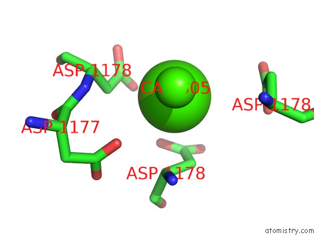
Mono view
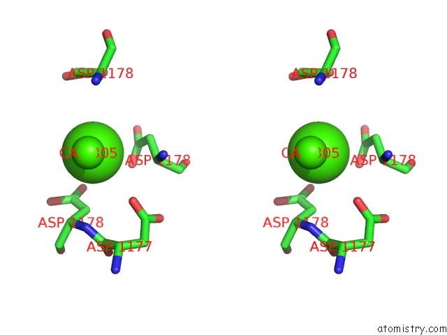
Stereo pair view
Mono view
Stereo pair view
A full contact list of Calcium with other atoms in the Ca binding
site number 1 of Structure of Cavab in Complex with Nimodipine within 5.0Å range:
|
Calcium binding site 2 out of 2 in 5kmf
Go back to
Calcium binding site 2 out
of 2 in the Structure of Cavab in Complex with Nimodipine
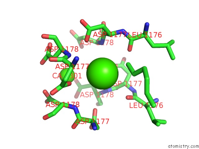
Mono view
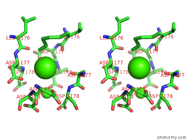
Stereo pair view
Mono view
Stereo pair view
A full contact list of Calcium with other atoms in the Ca binding
site number 2 of Structure of Cavab in Complex with Nimodipine within 5.0Å range:
|
Reference:
L.Tang,
T.M.El-Din,
T.M.Swanson,
D.C.Pryde,
T.Scheuer,
N.Zheng,
W.A.Catterall.
Structural Basis For Inhibition of A Voltage-Gated Ca(2+) Channel By Ca(2+) Antagonist Drugs. Nature V. 537 117 2016.
ISSN: ESSN 1476-4687
PubMed: 27556947
DOI: 10.1038/NATURE19102
Page generated: Wed Jul 9 07:28:36 2025
ISSN: ESSN 1476-4687
PubMed: 27556947
DOI: 10.1038/NATURE19102
Last articles
Zn in 3HTRZn in 3HT2
Zn in 3HTK
Zn in 3HRU
Zn in 3HSO
Zn in 3HSU
Zn in 3HSP
Zn in 3HSN
Zn in 3HR1
Zn in 3HS4