Calcium »
PDB 5m2s-5mi4 »
5m6d »
Calcium in PDB 5m6d: Streptococcus Pneumoniae Glyceraldehyde-3-Phosphate Dehydrogenase (Spgapdh) Crystal Structure
Protein crystallography data
The structure of Streptococcus Pneumoniae Glyceraldehyde-3-Phosphate Dehydrogenase (Spgapdh) Crystal Structure, PDB code: 5m6d
was solved by
C.Gaboriaud,
C.P.Moreau,
A.M.Di Guilmi,
with X-Ray Crystallography technique. A brief refinement statistics is given in the table below:
| Resolution Low / High (Å) | 40.52 / 2.00 |
| Space group | C 1 2 1 |
| Cell size a, b, c (Å), α, β, γ (°) | 81.041, 130.237, 79.837, 90.00, 119.64, 90.00 |
| R / Rfree (%) | 19.5 / 21.5 |
Other elements in 5m6d:
The structure of Streptococcus Pneumoniae Glyceraldehyde-3-Phosphate Dehydrogenase (Spgapdh) Crystal Structure also contains other interesting chemical elements:
| Chlorine | (Cl) | 2 atoms |
Calcium Binding Sites:
The binding sites of Calcium atom in the Streptococcus Pneumoniae Glyceraldehyde-3-Phosphate Dehydrogenase (Spgapdh) Crystal Structure
(pdb code 5m6d). This binding sites where shown within
5.0 Angstroms radius around Calcium atom.
In total only one binding site of Calcium was determined in the Streptococcus Pneumoniae Glyceraldehyde-3-Phosphate Dehydrogenase (Spgapdh) Crystal Structure, PDB code: 5m6d:
In total only one binding site of Calcium was determined in the Streptococcus Pneumoniae Glyceraldehyde-3-Phosphate Dehydrogenase (Spgapdh) Crystal Structure, PDB code: 5m6d:
Calcium binding site 1 out of 1 in 5m6d
Go back to
Calcium binding site 1 out
of 1 in the Streptococcus Pneumoniae Glyceraldehyde-3-Phosphate Dehydrogenase (Spgapdh) Crystal Structure
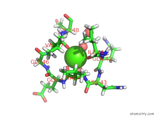
Mono view
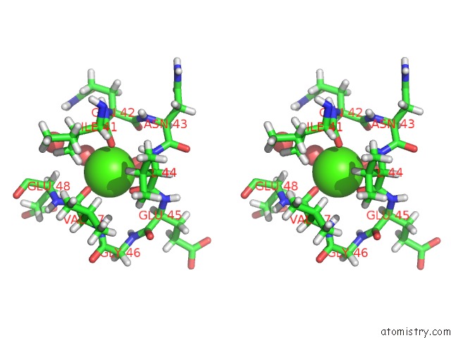
Stereo pair view
Mono view
Stereo pair view
A full contact list of Calcium with other atoms in the Ca binding
site number 1 of Streptococcus Pneumoniae Glyceraldehyde-3-Phosphate Dehydrogenase (Spgapdh) Crystal Structure within 5.0Å range:
|
Reference:
C.Moreau,
R.Terrasse,
N.M.Thielens,
T.Vernet,
C.Gaboriaud,
A.M.Di Guilmi.
Deciphering Key Residues Involved in the Virulence-Promoting Interactions Between Streptococcus Pneumoniae and Human Plasminogen. J. Biol. Chem. V. 292 2217 2017.
ISSN: ESSN 1083-351X
PubMed: 28011643
DOI: 10.1074/JBC.M116.764209
Page generated: Wed Jul 9 08:05:41 2025
ISSN: ESSN 1083-351X
PubMed: 28011643
DOI: 10.1074/JBC.M116.764209
Last articles
Fe in 2YXOFe in 2YRS
Fe in 2YXC
Fe in 2YNM
Fe in 2YVJ
Fe in 2YP1
Fe in 2YU2
Fe in 2YU1
Fe in 2YQB
Fe in 2YOO