Calcium »
PDB 5mi5-5mop »
5mon »
Calcium in PDB 5mon: Joint X-Ray/Neutron Structure of Cationic Trypsin in Complex with 2- Aminopyridine
Enzymatic activity of Joint X-Ray/Neutron Structure of Cationic Trypsin in Complex with 2- Aminopyridine
All present enzymatic activity of Joint X-Ray/Neutron Structure of Cationic Trypsin in Complex with 2- Aminopyridine:
3.4.21.4;
3.4.21.4;
Protein crystallography data
The structure of Joint X-Ray/Neutron Structure of Cationic Trypsin in Complex with 2- Aminopyridine, PDB code: 5mon
was solved by
J.Schiebel,
T.E.Schrader,
A.Ostermann,
A.Heine,
G.Klebe,
with X-Ray Crystallography technique. A brief refinement statistics is given in the table below:
| Resolution Low / High (Å) | N/A / 0.94 |
| Space group | P 21 21 21 |
| Cell size a, b, c (Å), α, β, γ (°) | 54.989, 58.537, 67.618, 90.00, 90.00, 90.00 |
| R / Rfree (%) | 17 / 18.1 |
Calcium Binding Sites:
The binding sites of Calcium atom in the Joint X-Ray/Neutron Structure of Cationic Trypsin in Complex with 2- Aminopyridine
(pdb code 5mon). This binding sites where shown within
5.0 Angstroms radius around Calcium atom.
In total only one binding site of Calcium was determined in the Joint X-Ray/Neutron Structure of Cationic Trypsin in Complex with 2- Aminopyridine, PDB code: 5mon:
In total only one binding site of Calcium was determined in the Joint X-Ray/Neutron Structure of Cationic Trypsin in Complex with 2- Aminopyridine, PDB code: 5mon:
Calcium binding site 1 out of 1 in 5mon
Go back to
Calcium binding site 1 out
of 1 in the Joint X-Ray/Neutron Structure of Cationic Trypsin in Complex with 2- Aminopyridine
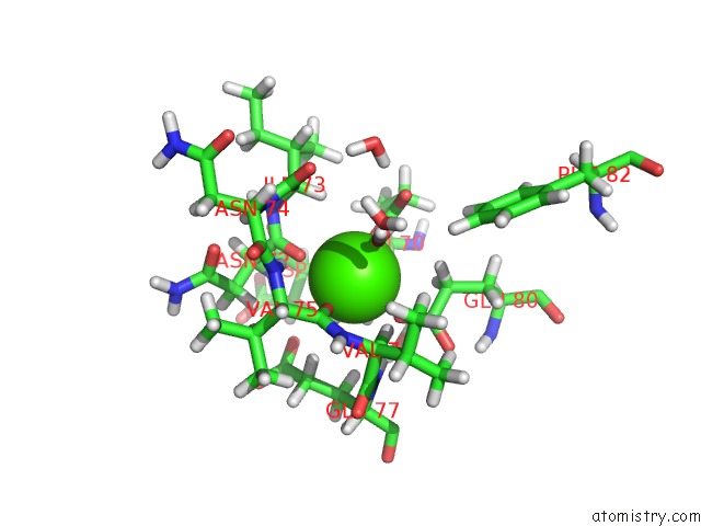
Mono view
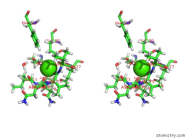
Stereo pair view
Mono view
Stereo pair view
A full contact list of Calcium with other atoms in the Ca binding
site number 1 of Joint X-Ray/Neutron Structure of Cationic Trypsin in Complex with 2- Aminopyridine within 5.0Å range:
|
Reference:
J.Schiebel,
R.Gaspari,
A.Sandner,
K.Ngo,
H.D.Gerber,
A.Cavalli,
A.Ostermann,
A.Heine,
G.Klebe.
Charges Shift Protonation: Neutron Diffraction Reveals That Aniline and 2-Aminopyridine Become Protonated Upon Binding to Trypsin. Angew. Chem. Int. Ed. Engl. V. 56 4887 2017.
ISSN: ESSN 1521-3773
PubMed: 28371253
DOI: 10.1002/ANIE.201701038
Page generated: Wed Jul 9 08:29:11 2025
ISSN: ESSN 1521-3773
PubMed: 28371253
DOI: 10.1002/ANIE.201701038
Last articles
Mg in 4LF7Mg in 4LF6
Mg in 4LF4
Mg in 4LF5
Mg in 4LCZ
Mg in 4LF2
Mg in 4LF1
Mg in 4LEM
Mg in 4LCK
Mg in 4LE0