Calcium »
PDB 5moq-5n2z »
5mst »
Calcium in PDB 5mst: Structure of the A Domain of Carboxylic Acid Reductase (Car) From Segniliparus Rugosus in Complex with Amp and A Co-Purified Carboxylic Acid
Protein crystallography data
The structure of Structure of the A Domain of Carboxylic Acid Reductase (Car) From Segniliparus Rugosus in Complex with Amp and A Co-Purified Carboxylic Acid, PDB code: 5mst
was solved by
D.Gahloth,
D.Leys,
with X-Ray Crystallography technique. A brief refinement statistics is given in the table below:
| Resolution Low / High (Å) | 113.64 / 1.72 |
| Space group | P 1 21 1 |
| Cell size a, b, c (Å), α, β, γ (°) | 69.300, 91.000, 113.780, 90.00, 92.84, 90.00 |
| R / Rfree (%) | 17.7 / 20.4 |
Calcium Binding Sites:
The binding sites of Calcium atom in the Structure of the A Domain of Carboxylic Acid Reductase (Car) From Segniliparus Rugosus in Complex with Amp and A Co-Purified Carboxylic Acid
(pdb code 5mst). This binding sites where shown within
5.0 Angstroms radius around Calcium atom.
In total 4 binding sites of Calcium where determined in the Structure of the A Domain of Carboxylic Acid Reductase (Car) From Segniliparus Rugosus in Complex with Amp and A Co-Purified Carboxylic Acid, PDB code: 5mst:
Jump to Calcium binding site number: 1; 2; 3; 4;
In total 4 binding sites of Calcium where determined in the Structure of the A Domain of Carboxylic Acid Reductase (Car) From Segniliparus Rugosus in Complex with Amp and A Co-Purified Carboxylic Acid, PDB code: 5mst:
Jump to Calcium binding site number: 1; 2; 3; 4;
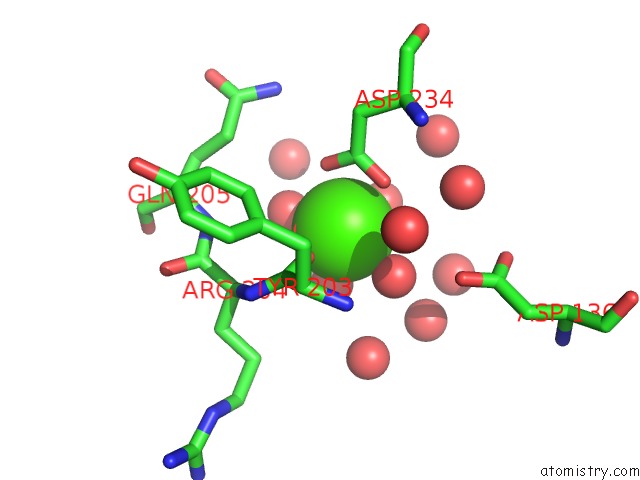
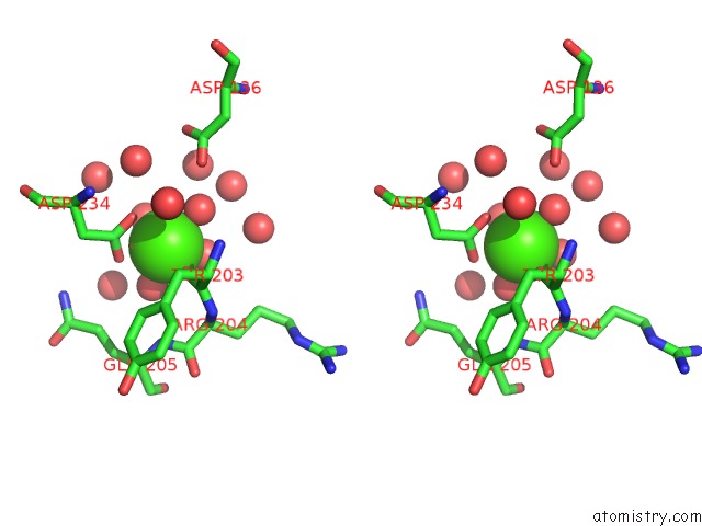
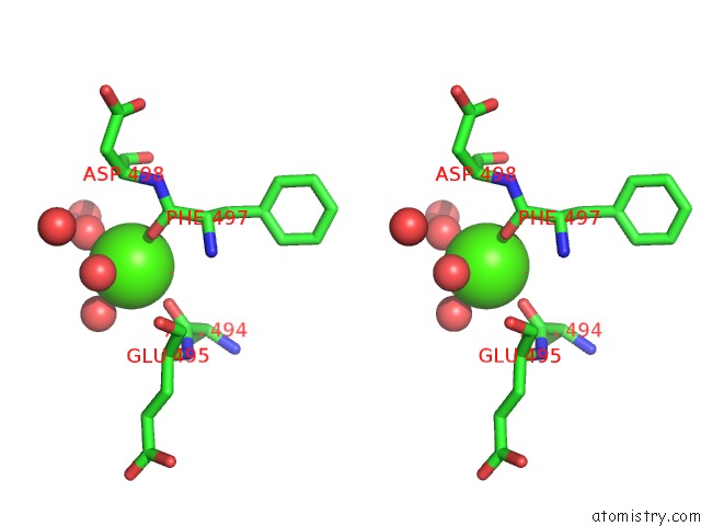
Calcium binding site 1 out of 4 in 5mst
Go back to
Calcium binding site 1 out
of 4 in the Structure of the A Domain of Carboxylic Acid Reductase (Car) From Segniliparus Rugosus in Complex with Amp and A Co-Purified Carboxylic Acid
Mono view
Stereo pair view
Mono view
Stereo pair view
A full contact list of Calcium with other atoms in the Ca binding
site number 1 of Structure of the A Domain of Carboxylic Acid Reductase (Car) From Segniliparus Rugosus in Complex with Amp and A Co-Purified Carboxylic Acid within 5.0Å range:
|
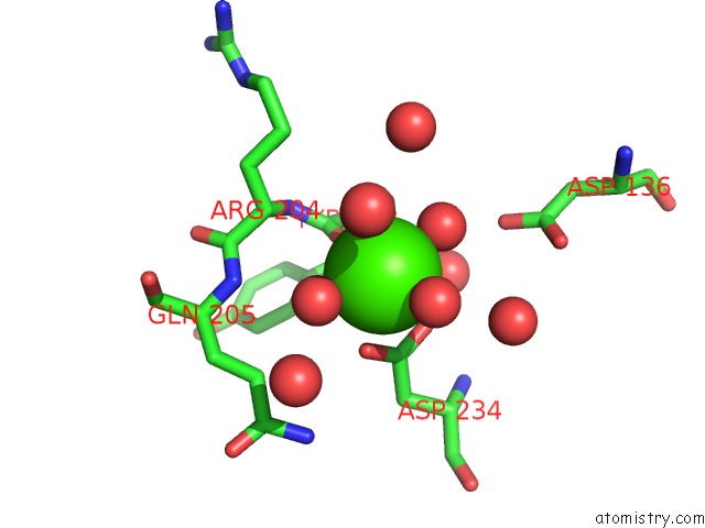
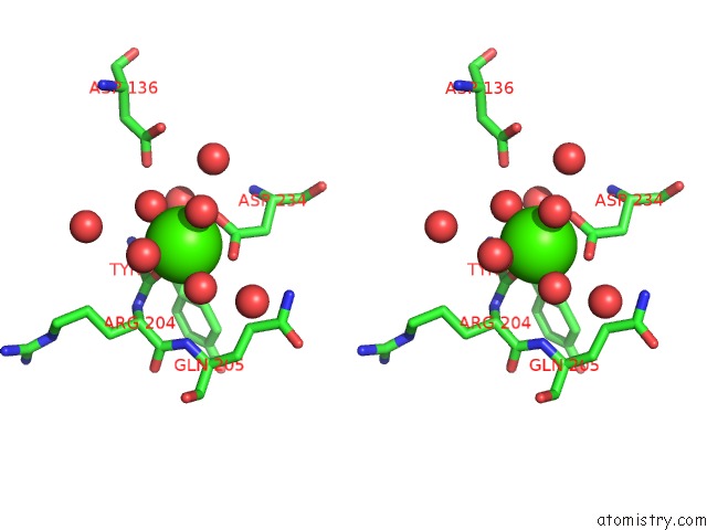
Calcium binding site 2 out of 4 in 5mst
Go back to
Calcium binding site 2 out
of 4 in the Structure of the A Domain of Carboxylic Acid Reductase (Car) From Segniliparus Rugosus in Complex with Amp and A Co-Purified Carboxylic Acid
Mono view

Stereo pair view
Mono view
Stereo pair view
A full contact list of Calcium with other atoms in the Ca binding
site number 2 of Structure of the A Domain of Carboxylic Acid Reductase (Car) From Segniliparus Rugosus in Complex with Amp and A Co-Purified Carboxylic Acid within 5.0Å range:
|
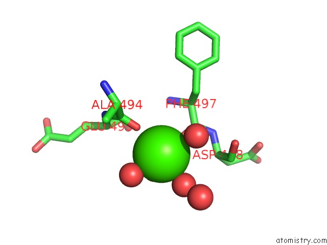
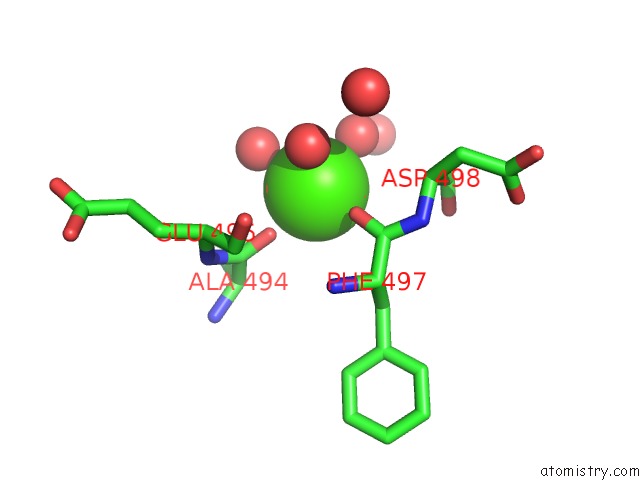
Calcium binding site 3 out of 4 in 5mst
Go back to
Calcium binding site 3 out
of 4 in the Structure of the A Domain of Carboxylic Acid Reductase (Car) From Segniliparus Rugosus in Complex with Amp and A Co-Purified Carboxylic Acid
Mono view
Stereo pair view
Mono view
Stereo pair view
A full contact list of Calcium with other atoms in the Ca binding
site number 3 of Structure of the A Domain of Carboxylic Acid Reductase (Car) From Segniliparus Rugosus in Complex with Amp and A Co-Purified Carboxylic Acid within 5.0Å range:
|
Calcium binding site 4 out of 4 in 5mst
Go back to
Calcium binding site 4 out
of 4 in the Structure of the A Domain of Carboxylic Acid Reductase (Car) From Segniliparus Rugosus in Complex with Amp and A Co-Purified Carboxylic Acid
Mono view
Stereo pair view
Mono view
Stereo pair view
A full contact list of Calcium with other atoms in the Ca binding
site number 4 of Structure of the A Domain of Carboxylic Acid Reductase (Car) From Segniliparus Rugosus in Complex with Amp and A Co-Purified Carboxylic Acid within 5.0Å range:
|
Reference:
D.Gahloth,
M.S.Dunstan,
D.Quaglia,
E.Klumbys,
M.P.Lockhart-Cairns,
A.M.Hill,
S.R.Derrington,
N.S.Scrutton,
N.J.Turner,
D.Leys.
Structures of Carboxylic Acid Reductase Reveal Domain Dynamics Underlying Catalysis. Nat. Chem. Biol. V. 13 975 2017.
ISSN: ESSN 1552-4469
PubMed: 28719588
DOI: 10.1038/NCHEMBIO.2434
Page generated: Wed Jul 9 08:31:12 2025
ISSN: ESSN 1552-4469
PubMed: 28719588
DOI: 10.1038/NCHEMBIO.2434
Last articles
Mg in 4X5CMg in 4X5E
Mg in 4X5V
Mg in 4X5B
Mg in 4X59
Mg in 4X58
Mg in 4X4V
Mg in 4X4S
Mg in 4X4R
Mg in 4X4Q